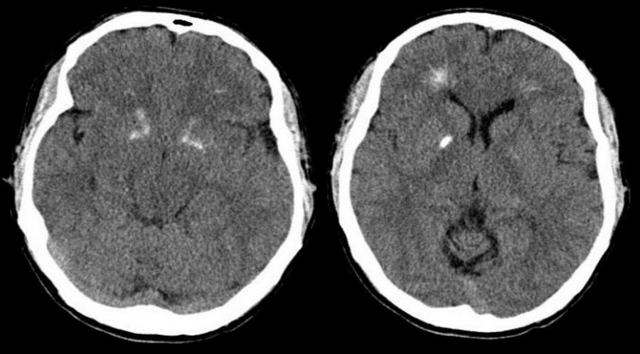
Dr. Allen Cherer is a neonatal care expert with over 30 years of medical accomplishments to his name.

Tag: treatment

dr-allen-cherer-blood

A Closer Look at Neonatal Polycythemia

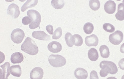
Polycythemia occurs when bone marrow is manufacturing too many red blood cells, thereby increasing the volume percentage of red blood cells in the blood. It can also be caused by a low plasma count in relation to blood cells, causing an imbalance. These excess cells, in turn, thicken the blood, which slows down the overall blood flow leading to more serious issues, such as blood clots. Polycythemia is essentially the opposite of anemia, which is when there are too few blood cells being produced. 

Neonatal is a term used to describe babies within the first 28 days of life. Neonatal polycythemia is diagnosed when a baby’s blood is composed of more than 65% red blood cells. This is a common problem with newborns, but that number is expected to decrease within a few hours if everything is normal. 

Some babies who are at a higher risk include those who are born small for their gestational age, babies who are post-term, infants of mothers with diabetes, twin-to-twin blood transfer in utero, low oxygen levels in fetal blood, and babies born with chromosomal abnormalities. 

It can be hard to diagnose this condition when babies are first born, because, aside from a high blood viscosity, babies might be asymptomatic before showing any metabolic changes due to this disorder. Samples should be taken from a largely free-flowing blood vessel in order to get an accurate hematocrit result. Signs that blood is affected include poor blood flow returning to a site after pressure is applied (peripheral perfusion), and blood having a ruddy, dusky appearance. There are also other clinical symptoms that include lethargy, irritability, tremors, seizures, lack of interest in feeding, hypoglycemia, rapid breathing (tachypnea), and bluish/grayish skin coloration (cyanosis).

There are several other conditions that share similar symptoms with neonatal polycythemia, which is why it is vital to rule them out before attempting any course of treatment. Some conditions that are similar include hypoglycemia, neological dysfunction, renal failure, or respiratory issues. Once a firm diagnosis is confirmed, there is a recommended course of action. 

Not all babies require treatment, but if there are signs of metabolic distress, the first priority is to lower the hematocrit by performing a partial exchange transfusion (PET). Either a saline solution may be used or a 5% protein solution. Saline is the preferred material because it won’t risk infection and has a better price point. It is strongly advised to avoid fresh frozen plasma because studies have shown a correlation between its use and necrotizing enterocolitis.

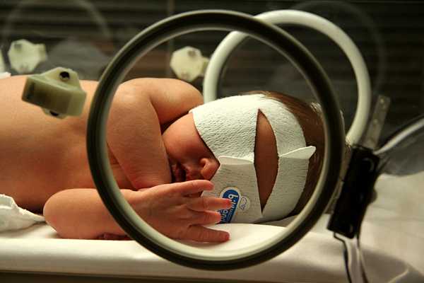
Jaundice Phototherapy

A Closer Look at Neonatal Jaundice

Neonatal jaundice is caused by unconjugated bilirubin in a newborn’s body. It’s presently the most common condition that requires medical attention or hospital readmission for newborns. This condition can have dire consequences, so early detection and treatment is crucial.

Symptoms Of Neonatal Jaundice

It’s common practice for hospitals to check newborns for jaundice before release,however parents should be aware of what to look for. Some symptoms of jaundice in infants include:

  • Yellow sclera (white part of the eyes)
  • Yellow skin, especially the abdomen and limbs
  • Baby is hard to awaken or lethargic
  • Baby is not gaining weight

If these symptoms appear after a child has been sent home from the hospital, it may be advisable to bring them back in for medical treatment.

Causes Of Neonatal Jaundice

Jaundice in infants can often be caused by the liver being underdeveloped. This leads to an inability for the liver to properly break down the bilirubin in their bodies. Besides immature livers, some other causes of this condition include:

  • Blood infection (sepsis)
  • Viral or bacterial infection
  • Malfunction of the liver
  • Deficient enzymes
  • Internal bleeding
  • Abnormal red blood cells

If symptoms of jaundice appear, the child will need to be checked for underlying causes to form a treatment plan.

Factors That Increase Risk Of Neonatal Jaundice

There are some factors that make jaundice more likely in infants. These factors include:

  • Bruising during birth
  • Mother and child’s blood types are different
  • Birth before 38 weeks
  • Breastfeeding difficulties

If a child has these risk factors, their parents and medical team will need to monitor them.

Treatment Of Neonatal Jaundice

To avoid acute or chronic complications, jaundice needs early treatment. Some ways it can be treated include:

  • Light therapy
  • Intravenous immunoglobulin
  • Exchange transfusion

These methods focus on diluting pathogenic antibodies in the child’s blood that cause jaundice or, in the case of light therapy, help the child’s body to more easily break down and excrete the excess bilirubin.


dr-allen-cherer-babyhead

A Closer Look at Neonatal Macrosomia

Neonatal macrosomia refers to babies weighing more than eight pounds and 13 ounces at birth. Approximately nine percent of infants are born with the condition. The larger the baby, the greater the risks to the mother and the infant. There are a variety of causes and risk factorsYour text to link… that lead to overweight newborns. Some causes are preventable.

Causes and Risks

  • Diabetes-Expectant mothers may have been diagnosed with diabetes before becoming pregnant. Others develop gestational diabetes during pregnancy. Blood sugars must be monitored and controlled otherwise, the infant develops with a larger amount of body fat.
  • Previous history-Women who have given birth to overly large infants in the past have a greater risk of having large babies in the future.
  • Obesity-There is a greater chance of having a baby with neonatal macrosomia if the mother is obese. Gaining too much weight during pregnancy also increases the risk.
  • Male infants-Neonatal macrosomia occurs more often in boy babies.
  • Overdue pregnancies-Pregnancies that extend two or more weeks beyond the estimated due date increase the chance that the infant will be overly large.
  • Mother’s age-Pregnant women over the age of 35 are more likely to have abnormally large babies.

Maternal Complications

  • Difficult labor-When an infant is too large, there is a likelihood that the baby becomes stuck in the birth canal, which may necessitate a C-section delivery.
  • Internal injuries-During the birthing process, the mother may suffer laceration or tearing of the vaginal tissues and perineal muscles.
  • Hemorrhaging-Internal injuries combined with the uterus’ inability to contract properly may lead to severe bleeding.
  • Uterine damage-Women who previously gave birth via C-section or had gynecological surgery have an increased risk of suffering from a uterine rupture.

Infant Complications

  • Hypoglycemia-Babies born with neonatal macrosomia have an increased risk of suffering from abnormally low blood sugars.
  • Obesity-Overly large infants are at a greater risk of becoming obese during childhood.
  • Metabolic syndrome-Neonatal macrosomia infants are likely to have metabolic syndrome. The condition is associated with hypertension, hyperglycemia, elevated cholesterol and excess body fat.

Prevention

Women must maintain a healthy weight before during and after pregnancy. While pregnant women should not gain ore than 35 pounds. Women diagnosed with diabetes must have their blood sugar continually monitored and controlled.

dr-allen-cherer-brain

DiGeorge Syndrome: An Overview

Primarily observed in children under the age of two, DiGeorge Syndrome is a rare neonatal chromosomal disorder affecting bodily development. The condition, which is also called 22q11.2 deletion syndrome, is caused by a defect in chromosome 22 and has a varying range of symptoms impacting both physical and mental growth. In some cases, it can be passed genetically from an affected parent to a child.

Due to the condition’s long list of symptoms — many of which are caused by a myriad of other conditions — DiGeorge Syndrome can be difficult to diagnose and treat, which means you will need to seek out an accurate and prompt evaluation from a trusted healthcare professional. Starting out, however, here is a quick overview of the condition to potentially point you in the right direction.

Knowing the signs

As mentioned before, symptoms of DiGeorge Syndrome can be vast, and therefore the condition can be hard to pinpoint at first. That said, there are several broad symptoms that have become listed as common warning signs; these include, but are certainly not limited to:

  • Cyanosis (a bluish tint to the skin caused by a lack of oxygen).
  • Learning difficulties, including those typically associated with Attention Deficit Disorder (ADD)
  • Skeletal abnormalities
  • Seizures and other epileptic symptoms
  • Feeding problems and failure to gain weight

Other, less externally evident symptoms may include autoimmune disorders, heart murmurs, frequent infections, and issues with the palate. In any scenario, the easiest and most established way to diagnose the condition is genetic testing, which can

Treating DiGeorge Syndrome

Unfortunately, there is currently no known cure for DiGeorge Syndrome — though certain symptoms may be individually treatable; this can fluctuate based on the urgency of the symptoms in question. For example, symptoms associated with certain immune-based disorders should ideally be addressed sooner than later to increase the chances of effective treatment. Other condition-based byproducts, like learning disabilities and anxiety, can be mitigated through proper intervention aimed at fostering intellectual growth and emotional stability.

These efforts may not treat the underlying root of the issue, but they can help on other fronts, such as improving the child’s overall quality of life.

Prevention can also be key in stopping the condition; if you feel you may have a family history of DiGeorge Syndrome, consult a specialist prior to any future pregnancies.

Providing Care for Drug-exposed Newborns: Time for the Next Step

During the years 1999-2013, the amount of prescription opioids dispensed in the United States nearly quadrupled, and since 2000, it is estimated that opioid use during pregnancy has tripled. Notably, the tragic consequences of the extreme availability of such drugs include abuse, physical dependence, and increasingly, death through inadvertent overdose.

newborn-boy-sleepingIn addition, for the individual pregnant woman, a minimum of two lives is affected: her own and that of her unborn child. The prevalence of prenatally exposed newborns to one or more illicit drugs approximates 6%. Neonatal Abstinence Syndrome (NAS) refers to the withdrawal symptoms from physical dependence experienced by the newborn exposed during pregnancy generally to illicit drugs, prescribed drugs, or to those opioids employed in medication-assisted treatment of maternal opioid addiction.

Withdrawal symptoms can vary markedly in terms of time of onset and severity but typically manifest as tremulousness, agitation, sleeplessness, and poor feeding. NAS increased threefold from 2000-2009 and frequently requires prolonged newborn hospitalization. It has been reported that aggregate hospital charges for NAS increased from 732 million dollars to 1.5 billion dollars with approximately 80% attributed to state Medicaid programs in 2012. Clearly, NAS is a costly public health problem resulting in significant human suffering and expense.

Traditionally, infants who are known to be at risk for NAS have been monitored in the postpartum unit after birth for at least 96 hours and withdrawal symptoms scored based on the Finnegan Scale developed in the mid 1970’s. Typically, if the scores exceed certain values, the newborn is admitted to a Special Care Unit where pharmacologic treatment is frequently started. As withdrawal symptoms subside, dosing is gradually tapered and ultimately stopped. The newborn is observed off medication and monitored for recurrence of disabling withdrawal symptoms. The entire process can generally result in a prolonged Special Care Unit hospital stay of 2-10 weeks.

With the seemingly overnight explosion in the number of newborns demonstrating withdrawal symptoms in the early 2000’s, medical caregivers and hospitals were caught off-guard. On short notice, staff addiction education, medication and weaning protocols, general care policies, and hospital space allocation were required. After a number of years of concerted, collaborative work, much has been learned and achieved in improving the care of the substance-exposed infant.

Nevertheless, pharmacologic treatment continues to require prolonged hospital stays, often in costly Special Care Units. In addition, it effectively excludes full participation by the eventual sole primary caregivers, ideally the parents. It is with these disturbing issues in mind that it is refreshing to note the work and studies over the past several years to further optimize the care provided to infants with NAS and their families.

One of the earlier studies to suggest the therapeutic benefits of a different approach to caring for the drug-exposed infant was that of Abrahams et al. published in the Canadian Family Physician in 2007. During the same period of frenzy involving inpatient hospital transfers, guaranteeing interobserver scoring reliability, pharmacologic treatment protocols, and nursing care directives, the Canadian group with extensive previous experience in addiction medicine reported in a retrospective cohort study the benefits of a rooming-in policy whereby infants remained with their mothers as primary caretakers.

They noted that infants who roomed-in were less likely to require pharmacologic therapy for withdrawal and more likely to be discharged to mother’s care compared to infant’s who received standard nursery care. Subsequently, other retrospective cohort studies both in Europe and the United States demonstrated equally beneficial effects of rooming-in regarding decreased requirement for pharmacologic therapy and decreased duration of hospital stay.

Most recently, the results of a quality collaborative project from the Children’s Hospital at Dartmouth Hitchcock were described in the May, 2016 Pediatrics and demonstrated the beneficial effects of combined standardized protocols and family-centered care in the management of the drug-exposed infant. Over time, the project safely reduced the number of infants requiring pharmacologic therapy, average length of stay, and overall hospital costs.

Among others, key drivers to success were prenatal education of family caregivers including expressed expectation that they would provide meaningful rooming-in care, baby-centered NAS scoring including on demand feeding schedules, pharmacologic therapy when necessary with dosing adjustment based on overall infant condition rather than solely Finnegan score and determined by a consistent team, and an infant “snuggler” volunteer program to assist families when times required their absence.

Overall, the project demonstrated that despite many practical obstacles to providing high quality care for drug-exposed newborns and their families in the hospital setting, where there’s a will, there’s a way.

Newborn Screening and Severe Combined Immune Deficiency

April (April 22-29) has been designated as National Primary Immunodeficiency Awareness Month and provides an opportunity to better understand the more than 250 rare, genetic disorders in which the body’s normal immune system is absent or functions improperly. Since an important function of the immune system is to protect against infection, patients with primary immune deficiency have an increased susceptibility to infection.

Severe Combined Immune Deficiency (SCID), popularized in the 1976 movie “The Boy in the Plastic Bubble”, is generally considered to be the most serious of the primary immunodeficiencies. There are at least 13 different genetic defects that can cause the disease; all of which are present at birth, involve missing T lymphocytes which are important in identifying and attacking perceived “invaders”, and affect the function of B lymphocytes which produce antibodies against infection.

The absence of T lymphocytes and antibody immunity results in severe infections, diarrhea, and failure to thrive. Regardless of the genetics, patients invariably succumb to an early death due to overwhelming infection. New approaches to diagnosis and management have changed what at one time was a dismal prognosis.

Treatment options have come a long way over the past 4 decades and include enzyme replacement, bone marrow transplant, and gene therapy. Paramount to this change is early diagnosis before the infant has had a chance to develop any serious infections.

The most effective therapy to date is immune reconstitution via stem cell transplant which has been shown to be highly successful (94%) if performed by 3.5 months of age. Hence, timing is crucial in terms of diagnosis and treatment.

Typically, infants with SCID appear totally normal at birth and have no family history of immunodeficiency. In the past, patients were primarily identified either by previous family history, physical manifestations, or after onset of life-threatening infection. Early identification of SCID has been achieved through the use of the 7-cell receptor excision circle (TREC) assay as part of the routine newborn screening program.

Absent or low TREC levels can indicate insufficient T lymphocyte production characteristic of SCID, as well as low T lymphocyte, non-SCID conditions as seen in DiGeorge Syndrome, Trisomy 21, CHARGE Syndrome, and ataxia telangiectasia. On May 21, 2010, The U.S. Department of Health and Human Services (HHS) recommended that every state include the assay as part of the newborn screen.

In the landmark study based on retrospective data on more than 3 million infants from 11 newborn screening programs using the TREC methodology conducted by Jennifer Puck, MD and colleagues and published August 20, 2014 in The Journal of the American Medical Association, the value of early detection and treatment of SCID was confirmed.

In addition, the study found an incidence rate almost twice as great (1 in 58,000 births) as had been previously estimated. Since the point of newborn screening is to identify conditions for which early treatment is life-saving, the study was a crucial step in the adoption of universal screening.

As of April 1, 2016, all, except for 11, states have adopted routine newborn screening programs for SCID. A 2016 study published in the Journal of Pediatrics by Ding and others provided an eloquent cost-benefit analysis of newborn screening in the treatment of Severe Combined Immune Deficiency.

Based on data obtained from 86,000 infants in Washington state, the study showed that newborn screening for SCID is clearly cost-effective. Hopefully, the study provides additional support in economic terms for the adoption of universal screening programs in all 50 states.

Physical Trauma and Pregnancy

Trauma is an important cause of maternal and fetal morbidity and mortality, and blunt abdominal trauma is a particular concern. Approximately 8% of pregnant women sustain some form of traumatic injury. Automobile accidents and falls account for most of the injuries.

Studies of pregnant women involved in automobile accidents have demonstrated increased rates of premature rupture of membranes, placental abruption, preterm birth, and stillbirth. A recent study examines pregnancies complicated by traumatic injuries and outcomes in relation to place of triage.

According to the retrospective study published in the Journal of the American College of Surgeons, pregnant women who sustained traumatic injuries and were triaged to trauma centers experienced improved pregnancy outcomes compared to women cared for in non-trauma hospitals. For the study, the researchers linked two databases, the Washington State Birth Events Records Database and the Comprehensive Hospital Abstract Recording System.

The method allowed them to assess the maternal and neonatal outcomes of all injured, pregnant women (3429 patients) who were hospitalized in Washington State between the years 1995 and 2012. The findings showed that after adjusting for a number of confounding factors, including injury severity score, pregnant women who were cared for in trauma facilities had better outcomes with significantly decreased odds of preterm labor, preterm birth, and low birth weight infants.

Since trauma centers are specialized medical facilities which have optimal processes and resources in place to monitor and treat injured patients, the study suggests that many injured, pregnant women may in fact be under-triaged and treated in non-trauma hospitals where their care may not be optimal.

According to the coauthors, John Distelhorst, DO, MPH and Vijay Krishnamoorthy, MD, the findings may lead to further analyses of state trauma systems and the triage of specific patient populations to improve quality of care and patient outcomes. To read more about this, please visit this site. 


Proton Radiotherapy: A Gentler Form of Radiation Treatment

Recent studies in Pediatric patients with brain tumors point to the efficacy of a new way of delivering radiation treatment which may result in improved long term outcomes for children.  Although not widely available, the new treatment, proton radiotherapy, focuses the radiation dose on the target area alone.  Standard photon (X-ray) radiation has the troublesome effect of exposing surrounding healthy tissues and organs  to the radiation as well. The new treatment has the distinct advantage of  getting  to “hard to get to” tumors.

A new study completed at the Massachusetts  General  Hospital  describes the results in a Pediatric patient population with medulloblastoma  treated with the usual combination of surgery, radiation, and chemotherapy.  Medulloblastoma is a fast-growing, high grade tumor always located in the cerebellum of the brain. It is a relatively rare tumor with more than 70% being diagnosed in children under 10 years of age. Like many tumors, its exact cause is unknown.  In  the study, the newer form of targeted radiation therapy was used and compared with the more  conventional  (photon) radiation. The results, as pointed out by Dr. Torunn Yock, Associate Professor of Radiation Oncology at Harvard Medical School, showed  comparable survival and tumor recurrence risks as well as long term hormone deficits between the two groups but far fewer side effects related  to hearing, cognition, and other organ systems.

The results are exciting in that they demonstrate the efficacy and safety  of proton radiotherapy with decreased long term side effects,  thus improving the quality of life in these young survivors.

Powered by WordPress & Theme by Anders Norén