Dr. Allen Cherer is a neonatal care expert with over 30 years of medical accomplishments to his name.

Tag: symptoms

dr-allen-cherer-blood

A Closer Look at Neonatal Polycythemia

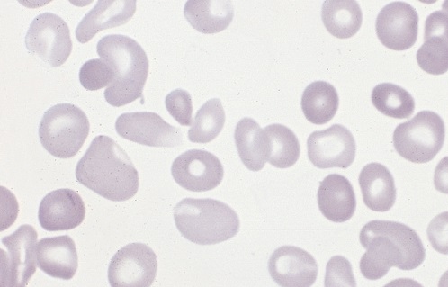
Polycythemia occurs when bone marrow is manufacturing too many red blood cells, thereby increasing the volume percentage of red blood cells in the blood. It can also be caused by a low plasma count in relation to blood cells, causing an imbalance. These excess cells, in turn, thicken the blood, which slows down the overall blood flow leading to more serious issues, such as blood clots. Polycythemia is essentially the opposite of anemia, which is when there are too few blood cells being produced. 

Neonatal is a term used to describe babies within the first 28 days of life. Neonatal polycythemia is diagnosed when a baby’s blood is composed of more than 65% red blood cells. This is a common problem with newborns, but that number is expected to decrease within a few hours if everything is normal. 

Some babies who are at a higher risk include those who are born small for their gestational age, babies who are post-term, infants of mothers with diabetes, twin-to-twin blood transfer in utero, low oxygen levels in fetal blood, and babies born with chromosomal abnormalities. 

It can be hard to diagnose this condition when babies are first born, because, aside from a high blood viscosity, babies might be asymptomatic before showing any metabolic changes due to this disorder. Samples should be taken from a largely free-flowing blood vessel in order to get an accurate hematocrit result. Signs that blood is affected include poor blood flow returning to a site after pressure is applied (peripheral perfusion), and blood having a ruddy, dusky appearance. There are also other clinical symptoms that include lethargy, irritability, tremors, seizures, lack of interest in feeding, hypoglycemia, rapid breathing (tachypnea), and bluish/grayish skin coloration (cyanosis).

There are several other conditions that share similar symptoms with neonatal polycythemia, which is why it is vital to rule them out before attempting any course of treatment. Some conditions that are similar include hypoglycemia, neological dysfunction, renal failure, or respiratory issues. Once a firm diagnosis is confirmed, there is a recommended course of action. 

Not all babies require treatment, but if there are signs of metabolic distress, the first priority is to lower the hematocrit by performing a partial exchange transfusion (PET). Either a saline solution may be used or a 5% protein solution. Saline is the preferred material because it won’t risk infection and has a better price point. It is strongly advised to avoid fresh frozen plasma because studies have shown a correlation between its use and necrotizing enterocolitis.

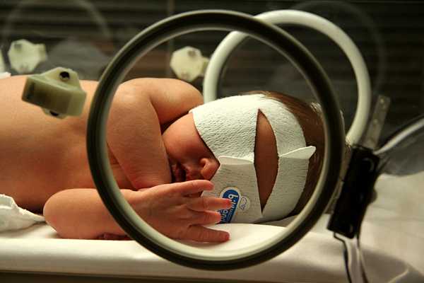
Jaundice Phototherapy

A Closer Look at Neonatal Jaundice

Neonatal jaundice is caused by unconjugated bilirubin in a newborn’s body. It’s presently the most common condition that requires medical attention or hospital readmission for newborns. This condition can have dire consequences, so early detection and treatment is crucial.

Symptoms Of Neonatal Jaundice

It’s common practice for hospitals to check newborns for jaundice before release,however parents should be aware of what to look for. Some symptoms of jaundice in infants include:

  • Yellow sclera (white part of the eyes)
  • Yellow skin, especially the abdomen and limbs
  • Baby is hard to awaken or lethargic
  • Baby is not gaining weight

If these symptoms appear after a child has been sent home from the hospital, it may be advisable to bring them back in for medical treatment.

Causes Of Neonatal Jaundice

Jaundice in infants can often be caused by the liver being underdeveloped. This leads to an inability for the liver to properly break down the bilirubin in their bodies. Besides immature livers, some other causes of this condition include:

  • Blood infection (sepsis)
  • Viral or bacterial infection
  • Malfunction of the liver
  • Deficient enzymes
  • Internal bleeding
  • Abnormal red blood cells

If symptoms of jaundice appear, the child will need to be checked for underlying causes to form a treatment plan.

Factors That Increase Risk Of Neonatal Jaundice

There are some factors that make jaundice more likely in infants. These factors include:

  • Bruising during birth
  • Mother and child’s blood types are different
  • Birth before 38 weeks
  • Breastfeeding difficulties

If a child has these risk factors, their parents and medical team will need to monitor them.

Treatment Of Neonatal Jaundice

To avoid acute or chronic complications, jaundice needs early treatment. Some ways it can be treated include:

  • Light therapy
  • Intravenous immunoglobulin
  • Exchange transfusion

These methods focus on diluting pathogenic antibodies in the child’s blood that cause jaundice or, in the case of light therapy, help the child’s body to more easily break down and excrete the excess bilirubin.


dr-allen-cherer-babyhead

A Closer Look at Neonatal Macrosomia

Neonatal macrosomia refers to babies weighing more than eight pounds and 13 ounces at birth. Approximately nine percent of infants are born with the condition. The larger the baby, the greater the risks to the mother and the infant. There are a variety of causes and risk factorsYour text to link… that lead to overweight newborns. Some causes are preventable.

Causes and Risks

  • Diabetes-Expectant mothers may have been diagnosed with diabetes before becoming pregnant. Others develop gestational diabetes during pregnancy. Blood sugars must be monitored and controlled otherwise, the infant develops with a larger amount of body fat.
  • Previous history-Women who have given birth to overly large infants in the past have a greater risk of having large babies in the future.
  • Obesity-There is a greater chance of having a baby with neonatal macrosomia if the mother is obese. Gaining too much weight during pregnancy also increases the risk.
  • Male infants-Neonatal macrosomia occurs more often in boy babies.
  • Overdue pregnancies-Pregnancies that extend two or more weeks beyond the estimated due date increase the chance that the infant will be overly large.
  • Mother’s age-Pregnant women over the age of 35 are more likely to have abnormally large babies.

Maternal Complications

  • Difficult labor-When an infant is too large, there is a likelihood that the baby becomes stuck in the birth canal, which may necessitate a C-section delivery.
  • Internal injuries-During the birthing process, the mother may suffer laceration or tearing of the vaginal tissues and perineal muscles.
  • Hemorrhaging-Internal injuries combined with the uterus’ inability to contract properly may lead to severe bleeding.
  • Uterine damage-Women who previously gave birth via C-section or had gynecological surgery have an increased risk of suffering from a uterine rupture.

Infant Complications

  • Hypoglycemia-Babies born with neonatal macrosomia have an increased risk of suffering from abnormally low blood sugars.
  • Obesity-Overly large infants are at a greater risk of becoming obese during childhood.
  • Metabolic syndrome-Neonatal macrosomia infants are likely to have metabolic syndrome. The condition is associated with hypertension, hyperglycemia, elevated cholesterol and excess body fat.

Prevention

Women must maintain a healthy weight before during and after pregnancy. While pregnant women should not gain ore than 35 pounds. Women diagnosed with diabetes must have their blood sugar continually monitored and controlled.

dr-allen-cherer-eyes

A Closer Look at Neonatal Alagille Syndrome

Roughly 1 out of 70,000 newborns is born with neonatal Alagille syndrome. This condition is often present at birth, though it can take several years for the symptoms to become present. Here are some important facts that parents need to know about neonatal Alagille syndrome.

What Is Neonatal Alagille Syndrome?

Neonatal Alagille Syndrome is a type of genetic disorder that children can inherit from their parents. People with neonatal Alagille syndrome have a mutated or missing copy of a gene labeled Jagged1 on chromosome 20. Problems with this gene are associated with a wide range of liver, heart, and eye problems. Due to a variety of skeletal abnormalities, people with neonatal Alagille syndrome tend to have a very characteristic appearance. Alagille syndrome is also referred to as Alagille-Watson syndrome, syndromic bile duct paucity, or arteriohepatic dysplasia.

What Are the Neonatal Alagille Syndrome Symptoms?

Not all people with neonatal Alagille syndrome have the same symptoms present. The condition can just cause minor issues like some changes to facial features, or it can cause severe heart and liver disease. Some of the most common symptoms include:

  • Deep-set eyes
  • Straight nose
  • Triangular face shape with pointed chin
  • Jaundice
  • Heart murmurs
  • Cardiac anomaly
  • Short height
  • Butterfly shaped vertebrae
  • Kidney abnormalities
  • Eye abnormalities

Parents should keep in mind that some symptoms, like kidney abnormalities, can take a while to notice. Signs that these organs are not functioning properly can include chronic itchiness, fatigue, pale skin, fluid retention, weakness, or abnormally pale feces.

How Is Neonatal Alagille Syndrome Treated?

Treatment for neonatal alagille syndrome typically relies on what symptoms the patient is presenting. Many children have mild enough Alagille syndrome that they just need to eat a healthy diet and get plenty of rest to help with issues like delayed puberty or failure to thrive. Certain medications can help with things like improving bile function, stabilizing kidney function, and helping the heart run properly. However, in more serious situations, surgery may be necessary. Surgeries can help to correct skeletal issues, replace malfunctioning kidneys and livers, or treat cardiac abnormalities.

dr-allen-cherer-syndrome

A Closer Look at Prader-Willi Syndrome

Prader-Willi syndrome is a genetic disorder that is generally rare. It occurs in approximately one out of every 12,000 births and results in weak muscle tone, delayed development, and poor growth. Around the age of two, children with the condition develop an insatiable appetite that can lead to obesity. The condition was first identified by Drs. Labhart, Prader, and Willi in 1965.

Symptoms

Symptoms of Prader-Willi syndrome include:

  • Weak muscle tone
  • Poor feeding in newborns
  • Delayed development
  • Obesity
  • Chronic overeating (hyperphagia)
  • Distinct facial features, which include almond-shaped eyes, turned-down mouth, narrowing of the forehead at the temples and a thin upper lip
  • Underdeveloped genitals
  • Compulsive behavior
  • Extreme stubbornness
  • Angry outbursts
  • Short stature
  • Delayed puberty
  • Sleep problems

Cause

Prader-Willi syndrome is caused by a loss of the function of the genes 11-13 on chromosome 15. In an estimated 70 to 80 percent of people with the syndrome, the region of genes is missing due to deletion of paternal genes. In other cases, there may be two copies of chromosome 15 inherited from the mother or a defect in the paternal genes on chromosome 15.

The loss of function on chromosome 15 disrupts the operation of a part of the brain known as the hypothalamus. The hypothalamus is responsible for the release of hormones in the body. Without the normal release of hormones, growth, body temperature, hunger, sleep, and sexual development can all be affected.

Complications

Obesity is common in those with Prader-Willi syndrome. People with the condition have lower muscle mass and are less likely to be physically active. As a result, they need fewer calories. Unfortunately, the hypothalamus is responsible for releasing hormones to signal feelings of fullness after eating. This results in people with the condition eating too many calories and becoming overweight or extremely obese.

Type 2 diabetes, sleep apnea, gallbladder, heart and liver problems may all result from obesity. People with the condition are encouraged to work with a dietitian to eat the proper amount of calories in order to prevent complications from occurring. Additionally, most people with Prader-Willi syndrome may experience fertility problems and are at an increased likelihood of developing osteoporosis.

Powered by WordPress & Theme by Anders Norén