Dr. Allen Cherer is a neonatal care expert with over 30 years of medical accomplishments to his name.

Category: Newborn Screening

dr-allen-cherer-babyhead

A Closer Look at Neonatal Macrosomia

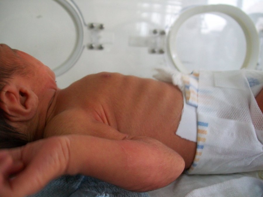
Neonatal macrosomia refers to babies weighing more than eight pounds and 13 ounces at birth. Approximately nine percent of infants are born with the condition. The larger the baby, the greater the risks to the mother and the infant. There are a variety of causes and risk factorsYour text to link… that lead to overweight newborns. Some causes are preventable.

Causes and Risks

  • Diabetes-Expectant mothers may have been diagnosed with diabetes before becoming pregnant. Others develop gestational diabetes during pregnancy. Blood sugars must be monitored and controlled otherwise, the infant develops with a larger amount of body fat.
  • Previous history-Women who have given birth to overly large infants in the past have a greater risk of having large babies in the future.
  • Obesity-There is a greater chance of having a baby with neonatal macrosomia if the mother is obese. Gaining too much weight during pregnancy also increases the risk.
  • Male infants-Neonatal macrosomia occurs more often in boy babies.
  • Overdue pregnancies-Pregnancies that extend two or more weeks beyond the estimated due date increase the chance that the infant will be overly large.
  • Mother’s age-Pregnant women over the age of 35 are more likely to have abnormally large babies.

Maternal Complications

  • Difficult labor-When an infant is too large, there is a likelihood that the baby becomes stuck in the birth canal, which may necessitate a C-section delivery.
  • Internal injuries-During the birthing process, the mother may suffer laceration or tearing of the vaginal tissues and perineal muscles.
  • Hemorrhaging-Internal injuries combined with the uterus’ inability to contract properly may lead to severe bleeding.
  • Uterine damage-Women who previously gave birth via C-section or had gynecological surgery have an increased risk of suffering from a uterine rupture.

Infant Complications

  • Hypoglycemia-Babies born with neonatal macrosomia have an increased risk of suffering from abnormally low blood sugars.
  • Obesity-Overly large infants are at a greater risk of becoming obese during childhood.
  • Metabolic syndrome-Neonatal macrosomia infants are likely to have metabolic syndrome. The condition is associated with hypertension, hyperglycemia, elevated cholesterol and excess body fat.

Prevention

Women must maintain a healthy weight before during and after pregnancy. While pregnant women should not gain ore than 35 pounds. Women diagnosed with diabetes must have their blood sugar continually monitored and controlled.

dr-allen-cherer-chest

Exploring Neonatal Sepsis

Neonatal Sepsis is a blood infection that infants may develop before reaching 90 days of age. Babies can also develop early-onset and late-onset sepsis.

Causes?

A bacteria named Eschericia coli (E coli) and Listeria can cause infants to develop sepsis. A specific streptococcus strain (Group B streptococcus or GBS) can also make an infant ill. If the baby’s mother contracts herpes simplex virus (HSV), this can also lead to neonatal sepsis.

An early-onset case usually develops 24 to 48 hours after the baby’s birth, usually by being exposed during birth. 

Contributors to early-onset sepsis:

  • Preterm delivery
  • GBS colonization during mother’s pregnancy
  • Placental tissues and amniotic fluid become infected (chorioamniontitis)
  • Early rupture of membranes (more than 18 hours)

Late-onset sepsis risks:

  • Extended hospitalization for infant
  • Keeping a catheter in baby’s blood vessel for an extended time

Symptoms?

  • Breathing problems
  • Changes in body temperature
  • Decreased bowel movements or diarrhea
  • Reduced movements
  • Low blood sugar
  • Reduced suckling
  • Heart rate is fast or slow
  • Seizures
  • Vomiting
  • Swollen abdomen
  • Jaundice (yellow skin and whites of eyes)

Diagnostic Tests?

Pediatricians perform the following diagnostic tests:

  • C-reactive protein
  • Blood culture
  • Complete blood count (CBC)
  • Lumbar puncture
  • Urine, skin or stool cultures to search for herpes virus
  • Chest X-ray (if baby has difficulty breathing)
  • Urine cultures

Treatments?

Even if the newborn is symptom-free, they will receive intravenous antibiotics. Babies younger than 4 weeks with fever or other symptoms receive IV antibiotics immediately.

The baby stays on antibiotics for three weeks if bacteria is in the spinal fluid or blood. This is shorter if no bacteria is present.

Acyclovir (antiviral medication) is given for HSV-caused infections.

If the baby has already gone home, it will be re-admitted to the hospital for treatment.

Outlook?

The infant may recover completely and show no evidence of any other problems. Neonatal sepsis can lead to infant death. The sooner treatment starts, the better the prognosis.

Potential Complications?

  • Disability after illness
  • Death

Prevention?

Pregnant mothers should receive preventive antibiotics if they have these illnesses:

  • Group B strep colonization
  • Chorioamnionitis
  • Has already had a baby with bacterial sepsis
  • This condition is preventable. Babies should be delivered 12 to 24 hours after water breaks.

Other Names?

Other names include:

  • Neonatal septicemia
  • Sepsis – infant
  • Sepsis neonatorium
dr-allen-cherer-eyes

A Closer Look at Neonatal Alagille Syndrome

Roughly 1 out of 70,000 newborns is born with neonatal Alagille syndrome. This condition is often present at birth, though it can take several years for the symptoms to become present. Here are some important facts that parents need to know about neonatal Alagille syndrome.

What Is Neonatal Alagille Syndrome?

Neonatal Alagille Syndrome is a type of genetic disorder that children can inherit from their parents. People with neonatal Alagille syndrome have a mutated or missing copy of a gene labeled Jagged1 on chromosome 20. Problems with this gene are associated with a wide range of liver, heart, and eye problems. Due to a variety of skeletal abnormalities, people with neonatal Alagille syndrome tend to have a very characteristic appearance. Alagille syndrome is also referred to as Alagille-Watson syndrome, syndromic bile duct paucity, or arteriohepatic dysplasia.

What Are the Neonatal Alagille Syndrome Symptoms?

Not all people with neonatal Alagille syndrome have the same symptoms present. The condition can just cause minor issues like some changes to facial features, or it can cause severe heart and liver disease. Some of the most common symptoms include:

  • Deep-set eyes
  • Straight nose
  • Triangular face shape with pointed chin
  • Jaundice
  • Heart murmurs
  • Cardiac anomaly
  • Short height
  • Butterfly shaped vertebrae
  • Kidney abnormalities
  • Eye abnormalities

Parents should keep in mind that some symptoms, like kidney abnormalities, can take a while to notice. Signs that these organs are not functioning properly can include chronic itchiness, fatigue, pale skin, fluid retention, weakness, or abnormally pale feces.

How Is Neonatal Alagille Syndrome Treated?

Treatment for neonatal alagille syndrome typically relies on what symptoms the patient is presenting. Many children have mild enough Alagille syndrome that they just need to eat a healthy diet and get plenty of rest to help with issues like delayed puberty or failure to thrive. Certain medications can help with things like improving bile function, stabilizing kidney function, and helping the heart run properly. However, in more serious situations, surgery may be necessary. Surgeries can help to correct skeletal issues, replace malfunctioning kidneys and livers, or treat cardiac abnormalities.

dr-allen-cherer-echogenic

Echogenic Bowel: An Overview

Though it is not an incredibly common diagnosis, there are still thousands of fetuses diagnosed with an echogenic bowel each year. Roughly 1.8 percent of all fetuses have echogenic bowel, and their parents often wonder what exactly this term means. Understanding all the details about echogenic bowel will help parents discover how to handle this condition.

What Is Echogenic Bowel?

This term is simply used to refer to a fetal bowel that is abnormally bright when viewed on an ultrasound. Ultrasounds are always in shades of black, grey, and white, with denser tissue being bright white and fluid being black. Usually, the bowels of the fetus tend to be a dark grey on an ultrasound because they are softened tissue, but if a fetus has echogenic bowel, their bowels may look as bright as thick bones like the pelvic bone.

What Does It Mean to Have Echogenic Bowel?

When parents first hear this word, they often start to wonder if it is a serious problem. The reality is that it is not always a sign of a health condition. 0.5 percent of all perfectly healthy fetuses have an echogenic bowel, and it can just occur due to various fluctuations in growth. However, echogenic bowel can be a cause for concern because it is more common in babies born with Down syndrome or cystic fibrosis. It can also be a sign that the fetus is suffering from an intestinal obstruction or an infection like cytomegalovirus or toxoplasmosis.

How Is Echogenic Bowel Treated?

The method for addressing echogenic bowel typically depends on the severity. Low grade echogenic bowels which are less dense than bone are normally harmless, so doctors tend to take a “wait and see” approach. They normally recommend taking a more detailed ultrasound in a few weeks to check up on the growth of the fetus. However, for more severely echogenic bowels, doctors may recommend a maternal serum screening, blood tests, or an amniocentesis to determine if the fetus has Down syndrome, infections, or cystic fibrosis. To make sure that the pregnancy continues safely, doctors will usually do regular fetal monitoring to make sure the fetus is growing properly following a display of echogenic bowel.

dr-allen-cherer-distress

Respiratory Distress in Newborns

Respiratory distress syndrome, or RDS, is a common lung complaint for infants. This is especially true in premature babies, born before 37 weeks. The more premature the baby, the greater the chance the child will develop RDS.

RDS is caused by a shortage of pulmonary surfactant. Surfactant is a liquid that helps keep air sacs in the lungs, known as alveoli, open. Alveoli are critical. They are the site of the exchange of oxygen and carbon dioxide. They make it possible for the blood to be oxygenated fully. Since surfactant makes this possible, it’s a very important substance indeed.

There are several risk factors for RDS. In addition to prematurity, babies with RDS are more likely to be white, male, and multiples. Mothers with diabetes are more likely to give birth to RDS babies. Babies delivered by c-section are also more likely to develop this problem.

Parents of babies with some of these risk factors should be aware of the symptoms of respiratory distress syndrome. Babies with RDS breathe fast. They may grunt, making an ugh sound with each breath. Their nostrils will flare every time they breathe. Finally, they can have retractions, where the skin pulls under the rib cage or in between ribs with each breath. Their skin may not be as pink as that of a typical baby.

Luckily, there is treatment for RDS these days. Delivery of oxygen by nasal cannula is one treatment. A CPAP, or continuous positive airway pressure, machine can be used to push air into an infant’s lungs. This will keep the alveoli open. Severe cases of RDS can require a ventilator.

Ventilators are a serious measure. They require intubation, or a tube being placed down the infant’s windpipe. Ventilators are only used in babies who can’t breathe well without assistance.

In addition to helping deliver more oxygen, the issue of a lack of surfactant can also be addressed. Surfactant can be delivered directly to the lungs, also via intubation. Medications to calm the infant are also used, especially when intubation is required.

RDS can sometimes also be associated with infections. In those cases, antibiotics may be given to the infant. Not every baby requires all of these treatments. In some cases, babies get worse before getting better. RDS is, in general, very treatable.

dr-allen-cherer-myths

Persistent Myths in Neonatology

Neonatology is a medical specialty that deals with the care of newborns. It’s a subspecialty of pediatrics. In recent years, neonatologists have taken steps to demystify infant care. Part of that means confronting myths about caring for babies that have existed for generations.

Typically, these myths are spread by well-meaning friends and families. They want to help new parents and truly believe they’re passing on sound advice. However, much of this advice is badly out of date. Evidence-based medicine supported by double-blind trials has disproved much of it.

Nutrition and diet are common areas where myths abound. There are several nutrition myths about preemies and full-term babies. Nutrition myths can be focused on what babies eat, how they are fed and how their meals are prepared.

An example of a nutrition myth is that infant formula must be sterilized before feedings. This hasn’t been a recommended practice since the 1950s. In developed countries with clean water, preparing bottles with clean tap water is sufficient. Bottles and nipples should, of course, be washed with soap and water. But boiling is not necessary.

Other myths that persist are often centered on fevers. Parents misunderstand which fevers are serious. They can also attribute fevers to milestones like teething. Studies since the 1990s have shown that there’s actually no link between fever and teething. Even on days when new teeth erupt, an average temperature should not be above 100 degrees. This is a slight elevation, but not a dangerous fever.

Parents typically see fevers of about 102 as serious, high fevers. This is rarely the case. While calling a pediatrician can be prudent in these cases, typically the fever itself is not serious. Causes of fever can also be misunderstood, even by medical professionals. Sometimes, doctors attribute fever in newborns to dehydration. While this may be the case, it’s also important to rule out causes like infection before settling on that diagnosis.

It’s important to continue to talk about these neonatology myths. They are often handed down from previous generations in families. While they represent advice that was cutting-edge in the past, they can create more work for parents in the present. Medical professionals should also ensure that they are keeping up with literature, so that they can avoid pitfalls in treating newborn babies.

dr-allen-cherer-newborn

Neonatology: an overview

Neonatology is a type of pediatrics, focusing specifically on medical care for newborns. The primary patients of neonatology are newborn infants who were born ill or became ill shortly after birth.

Here is a quick overview of this medical concentration, for those unfamiliar.

Origins of Neonatology

Neonatology is a very recent concentration of pediatrics. High infant mortality rates existed as early as the late 1800s. The first premature infant incubator station was created in Chicago by Joseph DeLee. The first NICU (newborn intensive care unit) was established in New Haven, Connecticut. Neonatology was officially recognized as an official subspecialty of pediatrics in 1975 by the American Board of Pediatrics.

Modern Neonatologists

Modern neonatology physicians are not here to help with minor problems; a normal pediatrician will be able to assist with most medical issues in infants. A neonatologist is trained to deal with high-risk situations. Premature babies, birth defects, and other serious issues are handled by neonatologists.

Neonatologists are serious doctors, and it takes serious time to become qualified. In addition to a standard college education, a doctor must have 4 years of medical school, 3 years of residency in pediatrics, 3 more years of residency in newborn intensive care, and they must be certified by the American Board of Pediatrics.

In addition to neonatologists, there are neonatal nurse practitioners. These nurses are specialized in neonatal care, and they will be assisting the physician along the way. They are able to diagnose some issues, prescribe medication, and some can even perform medical procedures themselves.

A neonatologist may assist with the diagnoses of breathing disorders, certain infections, and birth defects. They will also be the primary strategist in treatment options for an infant. They will formulate nutrition plans to make sure an infant will have maximum growth. A neonatologist will work closely with other medical staff, pediatricians, and nursing staff to assist with any serious illnesses in newborns.

Neonatologists are Best for Newborns

Minor problems for adults could mean possible death for an infant. That’s why specialists are needed for infants. There are also many common postpartum issues that a neonatologists can assist with. Many of these are routine for them; however, rare diseases and disorders can be diagnosed by a neonatologist as well.

Neonatal jaundice, neonatal cancer, inborn errors of metabolism, neonatal diabetes mellitus, neonatal herpes simplex, and neonatal seizure are a few of the more common problems a neonatologist will assist with.

2017 09 30 05 39 43 1100x733

The Prevention of Cystic Fibrosis in Newborns

Cystic fibrosis is one of the most common conditions caused by genetics. One baby out of every 3,500 live births will have cystic fibrosis. Cystic fibrosis affects the respiratory and digestive systems. Normally, the mucus that lines organs in the body is thin and slick with a consistency slightly thicker than water. Babies who are born with cystic fibrosis have mucus that is sticky and thick. If the mucus builds up, it makes breathing difficult. Additionally, the thick mucus can prevent nutrients from being absorbed properly, which may lead to poor growth.

Causes of Cystic Fibrosis

Cystic fibrosis is a genetic condition and must be inherited from a parent. A gene mutation causes cystic fibrosis. When it is passed on to a child, the baby will be born with the condition. There is no way to prevent cystic fibrosis from occurring in newborns.

Diagnosis of Cystic Fibrosis

In the United States, newborns are regularly screened for cystic fibrosis. A small amount of blood is taken from the newborn and examined for high levels of a chemical called immunoreactive trypsinogen (IRT). If IRT levels are higher than normal, a secondary test will be run in order to rule out other conditions that can also present with high IRT levels.

The second test is known as a “sweat test.” Newborns with cystic fibrosis have more salt in their sweat than normal. Medication will be administered to the baby that causes sweat to form. This sweat will then be tested for sodium levels. If sodium levels are high, cystic fibrosis is typically diagnosed. Additional tests, such as genetic tests, may also be performed to confirm the diagnosis.

Treatment of Cystic Fibrosis in Newborns

When diagnosed early, cystic fibrosis has a higher success rate of treatment. Prescription medications can help prevent infections from occurring, reduce lung damage and decrease inflammation. Physical therapy will help loosen the thick mucus and make it easier for babies to breathe. A special diet will help increase food absorption and help newborns with the condition grow and thrive.

Cystic fibrosis is a life-threatening condition that requires continual care. Though there is no way to prevent cystic fibrosis in newborns, medical advancements can help babies diagnosed with the condition live longer and healthier lives than ever before.

dr-allen-cherer-neonatology

Neonatology: a Brief History

Physicians and scientists began recognizing that premature or ill newborns required specialized care in the 1700s. However, it would be another century before a physician would take the first steps toward improving neonatal health. In the coming years, advancements in science and technology steadily enhanced the chances that preterm infants survived.

19th Century 

French obstetrician Etienne Stephane Tarnier recognized that premature infants were unable to maintain their body temperature. The physician invented the first incubator using a wooden box with a glass lid. The heat was provided by a hot water bottle. As a result, infant mortality decreased by 28 percent.

Pierre-Constant Budin trained under Dr. Tarnier and became a pioneer in neonatal nutrition during the late 1800s. Dr. Budin was aware of the risks of feeding newborns cow’s milk due to pathogens. He encouraged his new mothers to breastfeed. He was also responsible for introducing tube feeding for preemies who were unable to feed naturally.

By the early 1900s, Martin Couney, one of Dr. Budin’s students, improved upon Tarnier’s incubator design. However, the medical community was not accepting and the devices were not used in hospitals. In order to gain attention for the need, Dr. Couney began treating infants free of charge and demonstrated his invention at expositions and fairs.

20th Century 

For the most part, premature or ailing infants were not provided medical care. It was not until after World War II that the medical community recognized the need to offer specialized care. During this era, hospitals began developing “Special Care Baby Units” that eventually evolved into NICUs. Along with providing sufficient warmth, the units ensured that the infants received oxygen. There was also increasing awareness of an infant’s susceptibility to infection, which led to stringent hand washing.

Formulas for premature infants were introduced during this time. The formulas contained increased levels of calcium, phosphorus, sodium and protein. However, the high protein levels soon created a number of problems. As such, whey proteins were used.

Beginning in the 1960s, laboratory tests and values were established to monitor infant health. Physicians created a way to evaluate blood gases, bilirubin levels and liver function along with checking electrolytes, blood sugar and oxygen levels.

Advancements in knowledge and technology meant that infants born after 23 weeks of gestation had a survival rate of 33 percent. Infants born after 24 weeks had a survival rate of 66 percent. The survival rates continue growing each year.

Congenital Hypothyroidism and Newborn Screening

Congenital Hypothyroidism and Newborn Screening

Newborn screening for Congenital Hypothyroidism (CH) is a major public health achievement. Thyroid hormone is essential for the maturation of brain function and somatic growth, and its deficiency early in life can lead to mental retardation. For the fetus, maternal thyroid status is important during the first half of gestation; thereafter, the fetus’  hypothalamus-pituitary-thyroid axis is functional in the normal situation. For the hypothyroid newborn, it is well documented that provision of thyroid hormone is critical during the first weeks of life to avoid severe intellectual impairment. Notably, congenital hypothyroidism is considered one of the most common preventable causes  of mental retardation.

Studies showed that affected newborns were rarely identified during the first months of life and were often missed until 1-3 years of age. Congenital hypothyroidism  was found to be  an ideal candidate with the introduction of dried blood newborn screening by Dussault in Canada. With the development of increasingly sensitive assays to measure thyroid hormone (T4) and thyroid stimulating hormone (TSH) using a dried blood spot (DBS), newborn screening programs have developed throughout much of the world. In the 1980s, the incidence of CH in the United States was estimated to be 1:3000-1:4000. More recently, screening programs have reported an increased incidence of 1:1400-1:2800, most probably due to changes in screening strategies and the identification of milder cases.

Typically, newborn screening requires a heel stick blood specimen obtained at 48-72 hrs of life prior to an infant’s discharge from the hospital. Most current assays measure TSH alone as an indicator of thyroid function.  Results above established cutoff levels generally signify thyroid gland dysfunction and indicate further testing. Although most helpful in early identification of term newborns with anatomic or functional thyroid gland abnormalities, the screening does miss a percentage of newborns, for example those with central hypothyroidism due to hypothalamic-pituitary failure and the increasingly larger group of preterm  infants with congenital hypothyroidism who demonstrate delayed elevations in TSH. Numerous questions remain regarding the optimal timing of follow up laboratory studies and even treatment of certain types of newborn thyroid dysfunction.Nevertheless, newborn screening has proved invaluable for the great number of affected newborns.

The American Academy of Pediatrics recommends the measurement of TSH in all newborns with the goal that all infants with CH be identified by 2 weeks of age and that effective treatment with thyroid hormone replacement be started such that serum TSH levels less than 5 mIU/L be achieved within 4 weeks of diagnosis. Unfortunately, despite the significant successes following early identification and treatment of newborns with CH, obstacles persist in reaching the Academy’s goals. Screening programs continue to be plagued with the practical problems of screening all newborns, particularly those discharged home early who are lost to recall or lost to follow up altogether.  In addition, dried blood specimens are collected or processed improperly. Delays occur with recall of infants with abnormal results and with appropriate referrals for definitive treatment and management. A recent study conducted in Utah and reported at the 86th Annual Meeting of the American Thyroid Association highlights some of the problems which currently exist. After reviewing the TSH assays of 4394 children under 2 years of age, 48% of initial samples with elevated  levels (>20 mIU/L) were obtained after  the first 2 weeks of life, 15% of the initial abnormal TSH assays were not retested, and only 34% of those infants with initial elevated TSH assays achieved the goal of TSH < 5 mIU/L within 28 days of the initial assay.

The final message is that it is not enough to rely on the known efficacy of newborn screening for congenital hypothyroidism, but greater vigilance must be exercised to maximize its benefits in the lives of children.

Ehrenkranz J, Butler A, Snow G, Bach P. oral Abstract 19. The Diagnosis and Treatment of Congenital Hypothyroidism in Utah 2006-2015. Presented at: American Thyroid Association Annual Meeting; September 21-25, 2016; Denver, Colorado

Page 2 of 2

Powered by WordPress & Theme by Anders Norén