Dr. Allen Cherer is a neonatal care expert with over 30 years of medical accomplishments to his name.

Category: Child Health Care

dr-allen-cherer-brain

DiGeorge Syndrome: An Overview

Primarily observed in children under the age of two, DiGeorge Syndrome is a rare neonatal chromosomal disorder affecting bodily development. The condition, which is also called 22q11.2 deletion syndrome, is caused by a defect in chromosome 22 and has a varying range of symptoms impacting both physical and mental growth. In some cases, it can be passed genetically from an affected parent to a child.

Due to the condition’s long list of symptoms — many of which are caused by a myriad of other conditions — DiGeorge Syndrome can be difficult to diagnose and treat, which means you will need to seek out an accurate and prompt evaluation from a trusted healthcare professional. Starting out, however, here is a quick overview of the condition to potentially point you in the right direction.

Knowing the signs

As mentioned before, symptoms of DiGeorge Syndrome can be vast, and therefore the condition can be hard to pinpoint at first. That said, there are several broad symptoms that have become listed as common warning signs; these include, but are certainly not limited to:

  • Cyanosis (a bluish tint to the skin caused by a lack of oxygen).
  • Learning difficulties, including those typically associated with Attention Deficit Disorder (ADD)
  • Skeletal abnormalities
  • Seizures and other epileptic symptoms
  • Feeding problems and failure to gain weight

Other, less externally evident symptoms may include autoimmune disorders, heart murmurs, frequent infections, and issues with the palate. In any scenario, the easiest and most established way to diagnose the condition is genetic testing, which can

Treating DiGeorge Syndrome

Unfortunately, there is currently no known cure for DiGeorge Syndrome — though certain symptoms may be individually treatable; this can fluctuate based on the urgency of the symptoms in question. For example, symptoms associated with certain immune-based disorders should ideally be addressed sooner than later to increase the chances of effective treatment. Other condition-based byproducts, like learning disabilities and anxiety, can be mitigated through proper intervention aimed at fostering intellectual growth and emotional stability.

These efforts may not treat the underlying root of the issue, but they can help on other fronts, such as improving the child’s overall quality of life.

Prevention can also be key in stopping the condition; if you feel you may have a family history of DiGeorge Syndrome, consult a specialist prior to any future pregnancies.

vaccination herd immunity

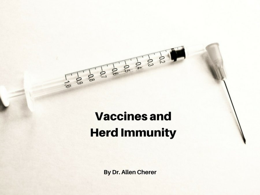
Vaccines and Herd Immunity

Herd immunity, also known as “community immunity”, is a phenomenon in which a group of people with high levels of immunity to a disease help prevent its spread.  So although individuals in a community can get infected, when a significant percentage is immune, they serve as a stopper on outbreaks and prevent them from spreading further.  Almost always, this is achieved through vaccination.  

Anti-vaccine activists dismiss the idea of herd immunity as not being real.  But that’s mostly because it’s another major benefit of vaccination, which goes against their overall agenda.  It also undercuts another favorite argument of the antivaxers, that vaccinated children have nothing to fear from unvaccinated children.  Of course, not all vaccines are 100% effective, yet another reason that herd immunity is so important.  

As the call against vaccines has become louder, we’ve noticed pockets of low vaccination in certain regions.  One place where it’s particularly caught on is Texas, where antivaxers have used pseudoscience and political concerns to get more people on board.  I recently read about one study from Stanford University that looked at what would happen if vaccine uptake declined nationwide.  For this study, they used publicly available data from the US Centers for Disease Control and Prevention to calculate the average rate of vaccination coverage for children ages 2-11 across the country.  With a mathematical model meant to calculate infectious disease transmission, they then estimated a distribution outbreak size related to a decrease in vaccine coverage.  

The model showed that a decrease in vaccination led to an increase in hypothetical measles cases.  The researchers found that a mere 5% decline in vaccine coverage for children would lead to a 3-fold increase in measles cases.  And this model doesn’t even account for all the vulnerable children, such as those too young to be vaccinated.  While this is of course a model, it’s not too much of a stretch to believe that even a small decrease in the number of vaccinated children could seriously damage the effectiveness of herd immunity.  In light of the measles outbreak currently sweeping through Europe, the need to not compromise herd immunity is more pressing than ever.  

Congenital Hypothyroidism and Newborn Screening

Congenital Hypothyroidism and Newborn Screening

Newborn screening for Congenital Hypothyroidism (CH) is a major public health achievement. Thyroid hormone is essential for the maturation of brain function and somatic growth, and its deficiency early in life can lead to mental retardation. For the fetus, maternal thyroid status is important during the first half of gestation; thereafter, the fetus’  hypothalamus-pituitary-thyroid axis is functional in the normal situation. For the hypothyroid newborn, it is well documented that provision of thyroid hormone is critical during the first weeks of life to avoid severe intellectual impairment. Notably, congenital hypothyroidism is considered one of the most common preventable causes  of mental retardation.

Studies showed that affected newborns were rarely identified during the first months of life and were often missed until 1-3 years of age. Congenital hypothyroidism  was found to be  an ideal candidate with the introduction of dried blood newborn screening by Dussault in Canada. With the development of increasingly sensitive assays to measure thyroid hormone (T4) and thyroid stimulating hormone (TSH) using a dried blood spot (DBS), newborn screening programs have developed throughout much of the world. In the 1980s, the incidence of CH in the United States was estimated to be 1:3000-1:4000. More recently, screening programs have reported an increased incidence of 1:1400-1:2800, most probably due to changes in screening strategies and the identification of milder cases.

Typically, newborn screening requires a heel stick blood specimen obtained at 48-72 hrs of life prior to an infant’s discharge from the hospital. Most current assays measure TSH alone as an indicator of thyroid function.  Results above established cutoff levels generally signify thyroid gland dysfunction and indicate further testing. Although most helpful in early identification of term newborns with anatomic or functional thyroid gland abnormalities, the screening does miss a percentage of newborns, for example those with central hypothyroidism due to hypothalamic-pituitary failure and the increasingly larger group of preterm  infants with congenital hypothyroidism who demonstrate delayed elevations in TSH. Numerous questions remain regarding the optimal timing of follow up laboratory studies and even treatment of certain types of newborn thyroid dysfunction.Nevertheless, newborn screening has proved invaluable for the great number of affected newborns.

The American Academy of Pediatrics recommends the measurement of TSH in all newborns with the goal that all infants with CH be identified by 2 weeks of age and that effective treatment with thyroid hormone replacement be started such that serum TSH levels less than 5 mIU/L be achieved within 4 weeks of diagnosis. Unfortunately, despite the significant successes following early identification and treatment of newborns with CH, obstacles persist in reaching the Academy’s goals. Screening programs continue to be plagued with the practical problems of screening all newborns, particularly those discharged home early who are lost to recall or lost to follow up altogether.  In addition, dried blood specimens are collected or processed improperly. Delays occur with recall of infants with abnormal results and with appropriate referrals for definitive treatment and management. A recent study conducted in Utah and reported at the 86th Annual Meeting of the American Thyroid Association highlights some of the problems which currently exist. After reviewing the TSH assays of 4394 children under 2 years of age, 48% of initial samples with elevated  levels (>20 mIU/L) were obtained after  the first 2 weeks of life, 15% of the initial abnormal TSH assays were not retested, and only 34% of those infants with initial elevated TSH assays achieved the goal of TSH < 5 mIU/L within 28 days of the initial assay.

The final message is that it is not enough to rely on the known efficacy of newborn screening for congenital hypothyroidism, but greater vigilance must be exercised to maximize its benefits in the lives of children.

Ehrenkranz J, Butler A, Snow G, Bach P. oral Abstract 19. The Diagnosis and Treatment of Congenital Hypothyroidism in Utah 2006-2015. Presented at: American Thyroid Association Annual Meeting; September 21-25, 2016; Denver, Colorado

Providing Care for Drug-exposed Newborns: Time for the Next Step

During the years 1999-2013, the amount of prescription opioids dispensed in the United States nearly quadrupled, and since 2000, it is estimated that opioid use during pregnancy has tripled. Notably, the tragic consequences of the extreme availability of such drugs include abuse, physical dependence, and increasingly, death through inadvertent overdose.

newborn-boy-sleepingIn addition, for the individual pregnant woman, a minimum of two lives is affected: her own and that of her unborn child. The prevalence of prenatally exposed newborns to one or more illicit drugs approximates 6%. Neonatal Abstinence Syndrome (NAS) refers to the withdrawal symptoms from physical dependence experienced by the newborn exposed during pregnancy generally to illicit drugs, prescribed drugs, or to those opioids employed in medication-assisted treatment of maternal opioid addiction.

Withdrawal symptoms can vary markedly in terms of time of onset and severity but typically manifest as tremulousness, agitation, sleeplessness, and poor feeding. NAS increased threefold from 2000-2009 and frequently requires prolonged newborn hospitalization. It has been reported that aggregate hospital charges for NAS increased from 732 million dollars to 1.5 billion dollars with approximately 80% attributed to state Medicaid programs in 2012. Clearly, NAS is a costly public health problem resulting in significant human suffering and expense.

Traditionally, infants who are known to be at risk for NAS have been monitored in the postpartum unit after birth for at least 96 hours and withdrawal symptoms scored based on the Finnegan Scale developed in the mid 1970’s. Typically, if the scores exceed certain values, the newborn is admitted to a Special Care Unit where pharmacologic treatment is frequently started. As withdrawal symptoms subside, dosing is gradually tapered and ultimately stopped. The newborn is observed off medication and monitored for recurrence of disabling withdrawal symptoms. The entire process can generally result in a prolonged Special Care Unit hospital stay of 2-10 weeks.

With the seemingly overnight explosion in the number of newborns demonstrating withdrawal symptoms in the early 2000’s, medical caregivers and hospitals were caught off-guard. On short notice, staff addiction education, medication and weaning protocols, general care policies, and hospital space allocation were required. After a number of years of concerted, collaborative work, much has been learned and achieved in improving the care of the substance-exposed infant.

Nevertheless, pharmacologic treatment continues to require prolonged hospital stays, often in costly Special Care Units. In addition, it effectively excludes full participation by the eventual sole primary caregivers, ideally the parents. It is with these disturbing issues in mind that it is refreshing to note the work and studies over the past several years to further optimize the care provided to infants with NAS and their families.

One of the earlier studies to suggest the therapeutic benefits of a different approach to caring for the drug-exposed infant was that of Abrahams et al. published in the Canadian Family Physician in 2007. During the same period of frenzy involving inpatient hospital transfers, guaranteeing interobserver scoring reliability, pharmacologic treatment protocols, and nursing care directives, the Canadian group with extensive previous experience in addiction medicine reported in a retrospective cohort study the benefits of a rooming-in policy whereby infants remained with their mothers as primary caretakers.

They noted that infants who roomed-in were less likely to require pharmacologic therapy for withdrawal and more likely to be discharged to mother’s care compared to infant’s who received standard nursery care. Subsequently, other retrospective cohort studies both in Europe and the United States demonstrated equally beneficial effects of rooming-in regarding decreased requirement for pharmacologic therapy and decreased duration of hospital stay.

Most recently, the results of a quality collaborative project from the Children’s Hospital at Dartmouth Hitchcock were described in the May, 2016 Pediatrics and demonstrated the beneficial effects of combined standardized protocols and family-centered care in the management of the drug-exposed infant. Over time, the project safely reduced the number of infants requiring pharmacologic therapy, average length of stay, and overall hospital costs.

Among others, key drivers to success were prenatal education of family caregivers including expressed expectation that they would provide meaningful rooming-in care, baby-centered NAS scoring including on demand feeding schedules, pharmacologic therapy when necessary with dosing adjustment based on overall infant condition rather than solely Finnegan score and determined by a consistent team, and an infant “snuggler” volunteer program to assist families when times required their absence.

Overall, the project demonstrated that despite many practical obstacles to providing high quality care for drug-exposed newborns and their families in the hospital setting, where there’s a will, there’s a way.

Antenatal Corticosteroid Use for Late Preterm Delivery

In 1972, Drs. Liggins and Howie published their landmark study demonstrating that antenatal corticosteroids administered to women 24-36 weeks of gestation reduced the incidence of respiratory distress syndrome and  neonatal mortality. Liggins had previously noted that lambs, treated with intrafetal  ACTH, cortisol, or dexamethasone, delivered prematurely, and sacrificed, demonstrated partially expanded  lungs.

Such alveolar stability was not typically noted until later in gestation. It suggested to Liggins that glucocorticoids might cause premature liberation of surfactant into the alveoli and served as the basis for his study. In the trial, the most significant difference in the incidence of respiratory distress syndrome among those treated vs. not treated with corticosteroids occurred in those gestations  less than 32 weeks.

Although those gestations treated between 32 and 37 weeks exhibited a decreased incidence of respiratory distress, the number did not reach statistical significance. Nevertheless, even at that time, Liggins postulated that mechanisms in addition to enhanced surfactant production and release might be responsible for the improved pulmonary function noted in more advanced gestations treated with antenatal corticosteroids.  

Interestingly, despite the findings of the initial study and similar results in multiple subsequent studies , the 1994 NIH Consensus report on the effect of corticosteroids for fetal maturation on perinatal outcomes found that only 20% of women who delivered newborns  501-1500 grams received the benefit of antenatal steroids. After a thorough review of available evidence, including  12 year neurodevelopmental follow up showing no adverse outcomes, the Consensus Panel felt  the benefits of antenatal administration of corticosteroids vastly outweigh the risks and all fetuses between 24 and 34 weeks gestation at risk of preterm delivery should be considered candidates for antenatal  treatment.

Only in those few pregnancies where corticosteroids would have an adverse effect on the mother or delivery was imminent  should steroid treatment be withheld. In addition, although Grade 1 evidence existed at the time to support the use of antenatal corticosteroids for gestations greater than 34 weeks, it was judged insufficient to recommend their use.

Since the Consensus statement, the use of antenatal corticosteroid use has become common and has resulted in considerable reduction in mortality and morbidity, as well as total health care costs. In addition, further neurodevelopmental follow up, including the original Auckland steroid trial participants, continues to demonstrate no adverse effects on psychological functioning and health-related quality of life. Other studies have demonstrated a decrease in overall respiratory disease in infants born beyond 34 weeks who had previously been exposed to antenatal corticosteroids when compared to unexposed infants born at similar gestations.

More than 300,000 pregnancies deliver in the late preterm period (34 0/7 – 36 6/7 weeks gestation) each year in the United States. Seventy per cent of Intensive Care Nursery admissions are late preterm newborns. Their increasing numbers and the broad range and severity of respiratory disorders with which they present beg for a re-evaluation of antenatal corticosteroid use in this range of gestations. This is especially appropriate with a better understanding of the multiple actions of corticosteroids as gestation approaches term.

A recent study, titled Antenatal Late Preterm Steroids (ALPS), a Randomized Trial to Reduce Neonatal Respiratory Morbidity, was published in The New England Journal of Medicine in April, 2016. The study enrolled over 2800 women with singleton pregnancies at high risk for late preterm delivery.

The participants were randomized to receive antenatal betamethasone by injection or a matching placebo. Greater than 80% of women in the trial delivered prior to 37 weeks gestation. The primary outcome was a neonatal composite of treatment in the first 72 hours (CPAP or High Flow Nasal Cannula for at least 2 hours, supplemental oxygen with fraction of inspired oxygen of at least 0.3 for at least 4 hours, mechanical ventilation, or ECMO) or stillbirth or neonatal death within 72 hours of birth.

The study found a significant decrease in neonatal respiratory complications in the group given the steroid treatment (11.6% vs. 14.4%). In addition, severe respiratory complications occurred significantly less frequently in the betamethasone group. The incidence of neonatal hypoglycemia  was increased in those treated with betamethasone (24% vs. 14.9%), but no other adverse neonatal outcomes were noted between the groups.

The study is authoritative due to its size, generalizability, and methodologic rigor. Although the issue of long term follow up cannot be specifically addressed, follow up studies of similar treatment in earlier gestations are reassuring. Late preterm births comprise a high risk group for hypoglycemia regardless of maternal antenatal steroid treatment and warrant vigilant monitoring during the newborn period.  In sum, the findings of the Antental Late Preterm Steroids study are consistent with other randomized controlled trials of antenatal corticosteroids administered at gestations less than 34 weeks.

Both the American College of Obstetrics and Gynecology with an endorsement by the American Academy of Pediatrics and the Society for Maternal-Fetal Medicine have addressed and published recommendations based on the study’s findings. Although the recommendations do not establish exclusive standards of care, the organizations approve the use of antenatal corticosteroids in certain defined late preterm pregnancies.  It is only with thoughtful application of the recommendations and further studies that the efficacy and safety of antenatal steroids in the late preterm pregnancy will be realized. It is a significant start.

Promoting Safe Sleep for Infants

Very few life events result in the anguish that comes with the death of an infant, especially one that is sudden and unexpected. Each year in the United States, approximately 3500 sudden, unexpected infant deaths (SUIDs) occur generally between the ages of 1 month and 1 year at a time when most infants sleep between 12 -18 hours/day.

They consist of three main types with Sudden Infant Death Syndrome (SIDS) being the predominant one, and deaths due to unknown causes and those due to accidental suffocation and strangulation in bed (ASSB) comprising the remainder.

The combined SUID death rate declined markedly following the 1992 American Academy of Pediatrics infant sleep recommendations and the initiation of the Back to Sleep campaign in 1994 with a primary focus on supine positioning during all infant sleep.

The combined SUID death rate decreased again slightly in 2009, and since that time has remained fairly constant. On the other hand, the ASSB, traditionally the least common of the three main causes of SUID, mortality rate remained unchanged until the late 1990s and has started a slow increase with its highest point in 2014.

Due in part to the success of the Back to Sleep campaign and to the increasing incidence of other sleep-related causes of SUID, the American Academy of Pediatrics broadened its focus since 2005 to include other factors resulting in an unsafe sleep environment and contributing to sleep-related infant deaths.

It is important to remember that the recommendations from the Safe to Sleep campaign are wholly derived from case-control studies and are based for the most part on epidemiologic studies including infants up to 1 year of age. The recommendations should therefore be applied to infants up to 1 year of age, except for those individuals in whom medical conditions warrant modification.

baby sleepingWhen it comes to safe sleep environment, remember the phrases “Back to Sleep”, “Bare is Best”, and “Room-sharing without Bed-sharing”. The basic underlying point to promote a safe sleep environment starts with every caretaker positioning every healthy infant on his or her back for every sleep.

Protective airway mechanisms prevent choking and aspiration. Only those infants with significant upper airway disorders warrant modification. Side sleeping is not recommended, and elevation while supine can be complicated by respiratory compromise if the infant’s position changes.

Preterm infants requiring prolonged hospitalization should also be maintained in the supine position during sleep when they are medically stable and long before they are ready for discharge to home.

Although the general recommendation pertains to infants up to one year of age, once an infant is capable of rolling from supine to prone and vice versa, the infant can remain in the sleep position that he or she assumes.
Since infants spend almost all of their time in a crib, bassinet, or play yard, these environments are especially important. Many infant deaths are associated with broken cribs with loose or missing parts.

Cribs should be no older than ten years and conform to the safety standards of the Consumer Product Safety Commission. Before use, the product should be checked for previous recall. Cribs require narrow slats and stable sides. Since 2011, federal safety standards prohibit the sale of drop side rail cribs. Specific mattresses designed for the crib should be firm and covered with a fitted sheet.

There should be no gaps larger than two finger breadths between the mattress and the crib. Soft materials or objects, such as pillows, comforters, or sheepskins even when covered with a sheet , should not be placed under a sleeping infant. Research shows that babies who sleep on soft surfaces which allow the baby’s head to sink into the surface are at higher risk for SIDS and suffocation.

If an infant falls asleep in a sitting device, such as a car safety seat, stroller, swing, or infant carrier, he or she should be removed from the product and moved to a crib or other appropriate firm flat surface as soon as practical.

When infant slings or cloth carriers are used, the infant’s head should be up and above the fabric, the face visible, and the nose and mouth not obstructed. The crib surface should be free of stuffed animals, pillows, toys, bumper pads, or blankets to reduce the risk of suffocation or entrapment. The crib, bassinet, or play yard should be positioned away from wall hangings, and the area should be free of blind and curtain cords which can result in strangulation.

Room-sharing without bed-sharing is recommended and is most likely to prevent accidental suffocation especially from overlaying, strangulation, and entrapment that might occur when an infant is sleeping in an adult bed. Soft mattresses, pillows, quilts, and loose bed linens provide a high risk environment for infants. Certainly, infants can be brought into the bed for feeding or comforting but should be returned to their own crib or bassinet when the parent is ready to return to sleep.

Epidemiologic studies have demonstrated increased risks for SIDS and suffocation when bed-sharing involves infants less than three months of age, other children or multiple persons, and caretakers who are excessively tired, current smokers, or are using medications or substances that impair alertness or ability to arouse. It is best to provide separate sleep areas and avoid co-bedding for twins and higher multiples in the hospital and at home.

Certain sudden, unexpected infant deaths are not preventable. Continuing research particularly related to SIDS will provide new insights into the mechanisms resulting in this tragedy. Nevertheless, vigilance in attending to those modifiable environmental risk factors is highly desirable.

Newborn Screening and Severe Combined Immune Deficiency

April (April 22-29) has been designated as National Primary Immunodeficiency Awareness Month and provides an opportunity to better understand the more than 250 rare, genetic disorders in which the body’s normal immune system is absent or functions improperly. Since an important function of the immune system is to protect against infection, patients with primary immune deficiency have an increased susceptibility to infection.

Severe Combined Immune Deficiency (SCID), popularized in the 1976 movie “The Boy in the Plastic Bubble”, is generally considered to be the most serious of the primary immunodeficiencies. There are at least 13 different genetic defects that can cause the disease; all of which are present at birth, involve missing T lymphocytes which are important in identifying and attacking perceived “invaders”, and affect the function of B lymphocytes which produce antibodies against infection.

The absence of T lymphocytes and antibody immunity results in severe infections, diarrhea, and failure to thrive. Regardless of the genetics, patients invariably succumb to an early death due to overwhelming infection. New approaches to diagnosis and management have changed what at one time was a dismal prognosis.

Treatment options have come a long way over the past 4 decades and include enzyme replacement, bone marrow transplant, and gene therapy. Paramount to this change is early diagnosis before the infant has had a chance to develop any serious infections.

The most effective therapy to date is immune reconstitution via stem cell transplant which has been shown to be highly successful (94%) if performed by 3.5 months of age. Hence, timing is crucial in terms of diagnosis and treatment.

Typically, infants with SCID appear totally normal at birth and have no family history of immunodeficiency. In the past, patients were primarily identified either by previous family history, physical manifestations, or after onset of life-threatening infection. Early identification of SCID has been achieved through the use of the 7-cell receptor excision circle (TREC) assay as part of the routine newborn screening program.

Absent or low TREC levels can indicate insufficient T lymphocyte production characteristic of SCID, as well as low T lymphocyte, non-SCID conditions as seen in DiGeorge Syndrome, Trisomy 21, CHARGE Syndrome, and ataxia telangiectasia. On May 21, 2010, The U.S. Department of Health and Human Services (HHS) recommended that every state include the assay as part of the newborn screen.

In the landmark study based on retrospective data on more than 3 million infants from 11 newborn screening programs using the TREC methodology conducted by Jennifer Puck, MD and colleagues and published August 20, 2014 in The Journal of the American Medical Association, the value of early detection and treatment of SCID was confirmed.

In addition, the study found an incidence rate almost twice as great (1 in 58,000 births) as had been previously estimated. Since the point of newborn screening is to identify conditions for which early treatment is life-saving, the study was a crucial step in the adoption of universal screening.

As of April 1, 2016, all, except for 11, states have adopted routine newborn screening programs for SCID. A 2016 study published in the Journal of Pediatrics by Ding and others provided an eloquent cost-benefit analysis of newborn screening in the treatment of Severe Combined Immune Deficiency.

Based on data obtained from 86,000 infants in Washington state, the study showed that newborn screening for SCID is clearly cost-effective. Hopefully, the study provides additional support in economic terms for the adoption of universal screening programs in all 50 states.

Reflections on Cesarean Section Rates

Cesarean section delivery is among the most common surgical procedures. It is estimated that in 2012 alone, over 22 million cesarean deliveries were performed worldwide. Data from the National Vital Statistics show that the total cesarean section rate in the U.S. in 1996 was approximately 21%.

Since that time, there has been a rapid increase in the rate, such that in 2011, close to 1 in 3 mothers delivered by cesarean. Although the rate has leveled since then, there remains no evidence that such a significant increase has been accompanied by a concomitant decrease in maternal or neonatal mortality.

Although cesarean delivery can be life-saving for the fetus, the mother, or both in certain cases, the concern exists that cesarean delivery is overused. Hence, the matter is a global health issue. Since one of the main driving forces for the increased total cesarean rate has been a marked shift to repeat cesarean delivery following a previous primary cesarean section, a concerted effort over the past several years has been to examine closely the factors related to the safe management of the nulliparous pregnancy.

As early as 1985, the World Health Organization (WHO) stated there was no justification for any region to have a cesarean delivery rate greater than 10-15 /100 live births. Nevertheless, the rates continued to increase worldwide with no scientific evidence indicative of substantial maternal or perinatal benefit.

In fact, a number of studies have associated higher rates of cesarean deliveries with negative consequences, including increased maternal and neonatal morbidity and mortality as well as increased consumption of limited health resources by procedures without medical indications.

In March 2014, a consensus report was issued by the American College of Obstetrics and Gynecology and the Society for Maternal-Fetal Medicine on the safe prevention of the primary cesarean section. Among other points, it addressed management guidelines for the most frequent indications for primary cesarean deliveries, namely, labor dystocia, abnormal or indeterminate fetal heart rate tracing, fetal malpresentation, multiple gestation, and suspected macrosomia.

The report encouraged obstetricians to allow more time to progress through a vaginal delivery without intervention, recommended improved and standardized fetal heart rate interpretation and management, and advocated access to non-medical interventionsduring labor, such as continuous labor and delivery support.

A study by researchers at Harvard Medical School and the Stanford University School of Medicine published December 2015 in The Journal of the American Medical Association suggested that based on analyses of cesarean section rates and maternal and neonatal outcomes among 194 WHO member countries the ideal rate of childbirth by cesarean section approximates 19% of all births as opposed tothe previously considered optimal rate of 10-15%. Although the finding is higher than the former target, it remains significantly lower than the current rate in U.S. hospitals.

In its April 2015 position statement on cesarean delivery, the WHO moved away from any target rate. Rather, it emphasized that every effort should be made to ensure cesarean sections are provided to the women in need and only be performed when medically necessary.

It is gratifying that the concept of “target rate” is no longer tied to the delivery of quality medical care. Primarily due to the lack of a consistent classification system to monitor and compare different obstetric profiles, meaningful data relative to cesarean section rates is missing.

It is only when such systems as the Robson Ten Group Classification System are widely adopted by institutions that valid “risk-adjusted”cesarean section rates can potentially be developed and comparisons be made between institutions, regions, and countries.

IVF and Childhood Cancer Risk

Assisted Reproductive Technology  (ART) has been successfully employed to treat infertility. In vitro fertilization (IVF)  is one form of ART, and it is unclear whether it increases the risks of birth defects . Since childhood cancers, such as leukemia, occur earlier in life, it is reasonable to question if factors related to early embryonic development and intrauterine environment may play a role in their occurrence.

A recent study from Norway suggests a slightly increased risk of childhood leukemia and Hodgkin’s lymphoma among children born through the use of ART. The findings were based on a review of children born between the years 1984 and 2011 and the use of ART. The data were then paired with cancer registry data. Out of 4500 cancer patients identified, 51 were conceived via ART.  Although overall cancer risk was not found to be increased among ART children, the odds of leukemia and Hodgkin’s lymphoma were.

As pointed out by Dr. Susan Amirian, assistant professor with the Baylor College of Medicine’s Duncan Cancer Center in an accompanying editorial, although the findings suggest a possible association, the very small numbers of actual cases  (17 cases of leukemia and 3 cases of Hodgkin’s lymphoma) call for caution in interpretation of the results. It would require more studies to establish a true association. Certainly other health factors that result in infertility may play a role in the apparent association.

At this point, the study results should not deter parents from using assisted reproductive technology and only careful monitoring of children born using the techniques is warranted. Take a look at this link for more information about this study.

Proton Radiotherapy: A Gentler Form of Radiation Treatment

Recent studies in Pediatric patients with brain tumors point to the efficacy of a new way of delivering radiation treatment which may result in improved long term outcomes for children.  Although not widely available, the new treatment, proton radiotherapy, focuses the radiation dose on the target area alone.  Standard photon (X-ray) radiation has the troublesome effect of exposing surrounding healthy tissues and organs  to the radiation as well. The new treatment has the distinct advantage of  getting  to “hard to get to” tumors.

A new study completed at the Massachusetts  General  Hospital  describes the results in a Pediatric patient population with medulloblastoma  treated with the usual combination of surgery, radiation, and chemotherapy.  Medulloblastoma is a fast-growing, high grade tumor always located in the cerebellum of the brain. It is a relatively rare tumor with more than 70% being diagnosed in children under 10 years of age. Like many tumors, its exact cause is unknown.  In  the study, the newer form of targeted radiation therapy was used and compared with the more  conventional  (photon) radiation. The results, as pointed out by Dr. Torunn Yock, Associate Professor of Radiation Oncology at Harvard Medical School, showed  comparable survival and tumor recurrence risks as well as long term hormone deficits between the two groups but far fewer side effects related  to hearing, cognition, and other organ systems.

The results are exciting in that they demonstrate the efficacy and safety  of proton radiotherapy with decreased long term side effects,  thus improving the quality of life in these young survivors.

Page 2 of 2

Powered by WordPress & Theme by Anders Norén