Dr. Allen Cherer | Neonatal Care & Pediatrics

Dr. Allen Cherer is a neonatal care expert with over 30 years of medical accomplishments to his name.

dr-allen-cherer-dna

DNA Sequencing Could Change How We Look at Genetic Neonatal Diseases

DNA sequencing is one of the most promising new technologies in terms of identifying the risk of disease, but it might not quite be ready for market. But regardless of concerns that DNA sequencing isn’t yet a safe screening method for newborn infants, chances are very strong that it will become a regular toolkit in preventative medicine sooner rather than later.

Routine blood tests are already part of standard procedure for infants born in the United States, and these tests can provide some substantive insight into potential future risks. But while a routine blood test can help identify dozens of different genetic conditions, that’s just scratching the surface of what can be accomplished with DNA sequencing. A study published in the American Journal of Human Genetics conducted DNA sequencing on 159 babies and found that 9% displayed anomalies that could predict genetic diseases that could appear in childhood. These include congenital heart disease and hearing loss.

But how much of an effect this testing could have on the health of infants is still an open question. Even co-author Alan Beggs questions how much substantive and actionable intelligence will arise from these genetic markers, at least for now. Nine percent is a low number, and many of these issues can be uncovered with the existing blood testing. Then there’s the fact that many of these genetic markers are not that well understood yet, and it can be difficult to understand how high of a risk such a genetic marker would actually pose.

Finally, there are a number of ethical and practical questions to consider. It can be hard to unpack issues of consent when dealing with the very genetic makeup of a child, and the rules behind the sharing of personal data, even as a means to better understand the map of human DNA, is still something like the Wild West. Finally, there are questions of how accessible this technology is and the costs associated for both medical providers and patients.

The running consensus now seems to be that DNA sequencing may be a beneficial choice in specific instances where parents are concerned about severe genetic disorders, but it’s not quite ready for primetime. As the technology and research continues to develop, it will likely become standard practice as a complement rather than a replacement for standard and accepted blood tests.

dr-allen-cherer-hepb

Fine-tuning the Elimination of Perinatal Hepatitis B Infection

Hepatitis B virus (HBV) infection  is a serious illness in the newborn and young infant.  The virus,  first discovered in the mid-1960s, is transmitted through percutaneous (i.e., puncture through skin) or mucosal (i.e., direct contact with mucous membranes) exposure to infectious blood or body fluids. The  virus is highly infectious, can be transmitted in the absence of visible blood, and remains viable on environmental surfaces for at least seven days.  Once the virus enters the body, it is transported to the liver where it replicates.  Although one generally thinks of the acute illness as a self-limited one in the adult with characteristic signs and symptoms, HBV infection in the infant is almost exclusively asymptomatic and hence, unrecognized. The devastating aspect of the infection is that the infant and young child frequently fail to clear the virus, and the illness becomes chronic. As many as 80-90% of infected infants progress to chronic infection, and chronically infected persons as adults are at increased risk of cirrhosis, hepatocellular carcinoma, and liver failure with approximately 25% dying from these serious complications.

Before 1982, an estimated 200,000-300,000 persons in the U.S. alone were infected with HBV annually, including approximately 20,000 infants. No effective pre-exposure prophylaxis existed, and only post-exposure prophylaxis in the form of hepatitis B immune globulin (HBIG) was available. However, the first hepatitis B vaccine was approved in the United States in 1981 and proved to be a real game changer. The availability of the vaccine set the stage for remarkable progress in the elimination of HBV infection among all age groups. With the advent of an effective vaccine, incurable hepatitis B infection had become preventable. The vaccine saves lives!

It is in this setting of disease prevention  through widespread vaccination that an evolving strategy to eliminate perinatal hepatitis B infection was initiated over 30 years ago. Early epidemiological studies had demonstrated that a major contributor to perinatal HBV infection is mother-to-child transmission  (MTCT) at the time of delivery. In utero infection is felt to account for less than 2% of infections. The risk of transmitting the virus was estimated to be 20-80% depending on the activity of the maternal infection. Initial attempts in the early 1980s to limit vertical HBV transmission were risk-based and aimed at identifying those pregnant women considered infectious by virtue of the serum marker, HBsAg. With reliable identification of mothers and expeditious treatment of their newborns with hepatitis B vaccine and HBIG, HBV infection could be prevented. However, it became clear within several years that such screening was inadequate with as many as 35-65% of HBsAg-positive women being missed. Consequently in 1988, the Advisory Committee on Immunization Practices (ACIP) of the Centers for Disease Control and Prevention recommended universal testing of all women early in each pregnancy such that at risk babies would receive appropriate post-exposure HBV prophylaxis. Throughout the 1990s, efforts were intensified to eliminate all HBV-related  illness through widespread vaccination of children, adolescents, and at-risk adults. Studies showed that receipt of the 3-dose hepatitis B vaccine series produced a protective antibody response in approximately 98% of healthy infants. During 1990-2004, the incidence of acute hepatitis B in the U.S. declined by 75%. The greatest decline (94%) occurred among children and adolescents, most likely due to increasing hepatitis B vaccine coverage. As of 2004, over 92% of children less than 3 years of age had been fully vaccinated with the complete series.

Coupled with the remarkable success of the hepatitis B elimination strategy is the knowledge that the task is not complete. As a  response to the persistence of perinatal HBV infection  and aware that errors in testing as well as in communication of results may occur, ACIP has recommended a change in the administration of the initial hepatitis B vaccine dose over time. Initially, the first dose could be administered to an infant born to a HBsAg-negative mother any time from birth to 2 months of age.  Subsequently the initial dose became the “birth dose” with the recommendation that it be given prior to discharge, and in 2017, the initial dose was to be administered within 24 hrs of birth. The previous  permissive language that allowed the dose to be delayed “on a case-by –case basis and only in rare circumstances” was omitted. Based on the fact that the vaccine alone is 75% effective in preventing MTCT, these changes reflect reality and provide basic protection. Then too, the emerging concept that maternal viral load (HBV DNA) plays a significant role in risk of MTCT now plays a prominent role in management.  Testing pregnant HBsAg-positive women for HBV DNA is now recommended to guide the use of antiviral therapy during the third trimester for the purpose of preventing perinatal HBV transmission.

stats photo dr. allen cherer

Fighting the P-Hack

It’s been said that statistics can be used to prove just about anything.  Take, for example, one study that I recently read about, which examined the link between vegetarianism among pregnant women and an increased risk of drug and alcohol abuse among their children.  The study examined over 5,000 women and their children, and finding that if their mothers ate little to no meat while pregnant, then the children were more likely to drink, smoke, and do drugs at 15.  It’s an interesting study, but at the same time, it’s one that could be part of a phenomenon that’s tragically common in the field of science, and is often used to push an agenda at the cost of objectivity.  I’m talking, of course, about “p-hacking”.  

In this phenomenon, “p” is the value used to determine statistical significance.  Ultimately, a difference between two groups is only meaningful if it’s statistically significant.  Let’s say a “p” value is less than .001: this means that there’s a less than 0.1% chance that an observed finding is due to chance, and more than a 99% chance that it represents a real difference.  This means that a statistically significant result is almost certainly real.  However, large datasets may involve countless variables.  If mining a dataset of 10,000 possible variables, for example, 10 statistically significant results should be treated as coincidence.  

Ultimately, p-hacking is a constant possibility when analyzing large datasets, so that treating every statistically significant result as real is dangerous.  Luckily, there are plenty of statistical tests to give a greater insight into whether or not a result is real.  The basic one is reproducibility.  If other data sets produce the same results, then you might just be onto something.  But until that happens, there’s no reason to believe that results are real.  Plenty of times, however, mining large datasets is used to find a point and push an agenda.  In the case of this study, eating meat.  Yet until that study is duplicated, you need to treat it with a grain of salt.  

hospital photo

Reducing Costs and Saving Lives

Sick newborns often rely on a ventilator to supply oxygen, and are tethered by a plastic endotracheal tube (ETT).  Often-times, this tube accidentally pops out.  This represents the fourth most common complication experienced by newborns in NICUs.  It can cause oxygen deficiency (hypoxia), high carbon dioxide levels in the blood, trauma to their airway, intraventricular hemorrhage, code events, and more.  “Unplanned extubations” also have the potential to nearly double the time of a hospital stay and increase the cost of care by $36,000 per patient.  Because of this, the Children’s National Health System’s NICU spearheaded a quality-improvement initiative to reduce the prevalence of unplanned extubations.

Many providers order a chest X-ray daily to verify the positioning of the ETT and prevent unplanned extubations.  However, this costs a lot of money, and potentially exposes infants to radiation.  Nobody needs to spend more money to expose themselves to more radiation, and that is particularly true for premature infants.  As part of their initiative, Children’s National has found that these same X-rays can be performed just twice weekly.  This sounds like a pretty simple initiative, but its consequences are surprisingly far-reaching: it lessens the chances of the breathing tube accidentally popping out, reducing radiation exposure, and saving roughly $1.6 million a year.  

The project started in July 2015, when Children’s National’s monthly X-ray expenditure was a whopping $289,520.  By the end of the year, that number had fallen down to $159,424, resulting in over $1 million in annual savings.  Within a month of implementation, unintended extubations dropped from 1.18 events per 100 ventilator days to .59.  Within five months, that number fell down .41.  Because of this initiative, unintended extubation rates at Children’s National are significantly lower than reported on various quality indices.  However, they don’t want to stop now, pledging to bring the rate down .3 events per 100 ventilator days.  I’m eager to see them reach their goal!

Pain in the NICU by dr. Allen Cherer

Pain in the NICU

When your child is placed in the Neonatal Intensive Care Unit (NICU), it’s an extremely stressful situation for everybody involved.  For parents, the stress of having their newborn in ICU is unimaginable.  But for the babies, maternal separation, noise, bright lights, procedures, and plenty of other operations make the experience overwhelming and extremely stressful in its own way, which can lead to various long-term consequences.  I recently read a very interesting blog post discussing why, and more importantly how, to deal with pain and stress in the NICU.    

It’s hard to imagine, but it was once believed that infants couldn’t feel pain.  Of course, we know now that that is hardly the case.  Although preterm infants have less localized and mature pain responses than their older counterparts, they still experience pain.  They often experience exaggerated pain responses, allodynia (pain from things that aren’t normally painful), and a longer latency in their responses.  The types of pain they experience include acute, postoperative, inflammatory, chronic, and visceral.  Pain experienced in these first stages of an infant’s life has potentially disastrous long-term consequences.  It may affect long-term memory, pain perception and responses, and possible alter social and cognitive development.  This is why understanding and addressing pain and stress in the NICU is so important.  

Stress and chronic pain is particularly difficult to assess, and is frequently superimposed by other types of pains.  When addressing acute pain due to procedures, it’s important to look at the procedure itself to see if it’s necessary, and also if you have access to local anesthetics.  Various sedatives, such as morphine, can be used for serious procedures, but each of these comes with its own set of long-term risks.  

Because of the stress and pain that comes with procedures, there’s been a movement to try and  limit the number of painful and stressful procedures.  There’s also been an effort to involve parents more to make the NICU environment less stressful.  This can involve swaddling, pacifiers, music, and family-centered care, all of which can reduce both stress and pain.  Because of this, parental participation, when combined with other methods, may be one of the safest options for the infant.  

vaccination herd immunity

Vaccines and Herd Immunity

Herd immunity, also known as “community immunity”, is a phenomenon in which a group of people with high levels of immunity to a disease help prevent its spread.  So although individuals in a community can get infected, when a significant percentage is immune, they serve as a stopper on outbreaks and prevent them from spreading further.  Almost always, this is achieved through vaccination.  

Anti-vaccine activists dismiss the idea of herd immunity as not being real.  But that’s mostly because it’s another major benefit of vaccination, which goes against their overall agenda.  It also undercuts another favorite argument of the antivaxers, that vaccinated children have nothing to fear from unvaccinated children.  Of course, not all vaccines are 100% effective, yet another reason that herd immunity is so important.  

As the call against vaccines has become louder, we’ve noticed pockets of low vaccination in certain regions.  One place where it’s particularly caught on is Texas, where antivaxers have used pseudoscience and political concerns to get more people on board.  I recently read about one study from Stanford University that looked at what would happen if vaccine uptake declined nationwide.  For this study, they used publicly available data from the US Centers for Disease Control and Prevention to calculate the average rate of vaccination coverage for children ages 2-11 across the country.  With a mathematical model meant to calculate infectious disease transmission, they then estimated a distribution outbreak size related to a decrease in vaccine coverage.  

The model showed that a decrease in vaccination led to an increase in hypothetical measles cases.  The researchers found that a mere 5% decline in vaccine coverage for children would lead to a 3-fold increase in measles cases.  And this model doesn’t even account for all the vulnerable children, such as those too young to be vaccinated.  While this is of course a model, it’s not too much of a stretch to believe that even a small decrease in the number of vaccinated children could seriously damage the effectiveness of herd immunity.  In light of the measles outbreak currently sweeping through Europe, the need to not compromise herd immunity is more pressing than ever.  

Congenital Hypothyroidism and Newborn Screening

Congenital Hypothyroidism and Newborn Screening

Newborn screening for Congenital Hypothyroidism (CH) is a major public health achievement. Thyroid hormone is essential for the maturation of brain function and somatic growth, and its deficiency early in life can lead to mental retardation. For the fetus, maternal thyroid status is important during the first half of gestation; thereafter, the fetus’  hypothalamus-pituitary-thyroid axis is functional in the normal situation. For the hypothyroid newborn, it is well documented that provision of thyroid hormone is critical during the first weeks of life to avoid severe intellectual impairment. Notably, congenital hypothyroidism is considered one of the most common preventable causes  of mental retardation.

Studies showed that affected newborns were rarely identified during the first months of life and were often missed until 1-3 years of age. Congenital hypothyroidism  was found to be  an ideal candidate with the introduction of dried blood newborn screening by Dussault in Canada. With the development of increasingly sensitive assays to measure thyroid hormone (T4) and thyroid stimulating hormone (TSH) using a dried blood spot (DBS), newborn screening programs have developed throughout much of the world. In the 1980s, the incidence of CH in the United States was estimated to be 1:3000-1:4000. More recently, screening programs have reported an increased incidence of 1:1400-1:2800, most probably due to changes in screening strategies and the identification of milder cases.

Typically, newborn screening requires a heel stick blood specimen obtained at 48-72 hrs of life prior to an infant’s discharge from the hospital. Most current assays measure TSH alone as an indicator of thyroid function.  Results above established cutoff levels generally signify thyroid gland dysfunction and indicate further testing. Although most helpful in early identification of term newborns with anatomic or functional thyroid gland abnormalities, the screening does miss a percentage of newborns, for example those with central hypothyroidism due to hypothalamic-pituitary failure and the increasingly larger group of preterm  infants with congenital hypothyroidism who demonstrate delayed elevations in TSH. Numerous questions remain regarding the optimal timing of follow up laboratory studies and even treatment of certain types of newborn thyroid dysfunction.Nevertheless, newborn screening has proved invaluable for the great number of affected newborns.

The American Academy of Pediatrics recommends the measurement of TSH in all newborns with the goal that all infants with CH be identified by 2 weeks of age and that effective treatment with thyroid hormone replacement be started such that serum TSH levels less than 5 mIU/L be achieved within 4 weeks of diagnosis. Unfortunately, despite the significant successes following early identification and treatment of newborns with CH, obstacles persist in reaching the Academy’s goals. Screening programs continue to be plagued with the practical problems of screening all newborns, particularly those discharged home early who are lost to recall or lost to follow up altogether.  In addition, dried blood specimens are collected or processed improperly. Delays occur with recall of infants with abnormal results and with appropriate referrals for definitive treatment and management. A recent study conducted in Utah and reported at the 86th Annual Meeting of the American Thyroid Association highlights some of the problems which currently exist. After reviewing the TSH assays of 4394 children under 2 years of age, 48% of initial samples with elevated  levels (>20 mIU/L) were obtained after  the first 2 weeks of life, 15% of the initial abnormal TSH assays were not retested, and only 34% of those infants with initial elevated TSH assays achieved the goal of TSH < 5 mIU/L within 28 days of the initial assay.

The final message is that it is not enough to rely on the known efficacy of newborn screening for congenital hypothyroidism, but greater vigilance must be exercised to maximize its benefits in the lives of children.

Ehrenkranz J, Butler A, Snow G, Bach P. oral Abstract 19. The Diagnosis and Treatment of Congenital Hypothyroidism in Utah 2006-2015. Presented at: American Thyroid Association Annual Meeting; September 21-25, 2016; Denver, Colorado

Noting the Extraordinary Success of Hib Vaccination

August is observed as National Immunization Awareness Month and is a time to highlight the extreme importance and value of vaccination for people of all ages. Vaccination serves as one of the best ways to protect infants, children, and adolescents from sixteen potentially harmful, and even deadly, diseases. Although it is common to think of the vaccines against measles, pertussis, and polio, an astonishingly important vaccine since the end of the 20th century has targeted the bacteria, Haemophilus influenzae type b (Hib).

Haemophilus influenzae is a small, pleomorphic, gram negative coccobacillus. Some strains of H. influenzae possess a polysaccharide capsule, and these strains are serotyped into six different types (a-f) based on their biochemically different capsules.

The H. influenzae strains with no capsule are termed nonencapsulated H. influenzae or nontypable H. influenzae (NTHi). H. influenzae type b is the most virulent, with its polysaccharide capsule being the main factor. Antibody to the capsule is the primary contributor to serum bactericidal activity, and increasing levels of antibody are associated with decreasing risk of invasive H. influenzae disease.

H. influenzae type b most commonly causes pneumonia, bacteremia, meningitis, epiglottitis, and cellulitis. Non-type b encapsulated forms present in a similar manner to type b infections, while non typable strains more commonly cause infections of the respiratory tract, such as pneumonia, otitis media, sinusitis, and conjunctivitis.

Generally, the mode of transmission is person to person by inhalation of respiratory tract droplets or by direct contact with respiratory tract secretions. Pharyngeal colonization by H. influenzae is relatively common, especially with nontypable and non-type b capsular strains.
Before effective Hib conjugate vaccines for infants older than 2 months were available in 1990, Haemophilus influenzae type b was the leading cause of invasive bacterial disease among children in the United States.

One in 200 children developed invasive Hib disease by 5 years of age; approximately 60% of these children had meningitis and 3-6% died from the disease. Of the Hib meningitis survivors, many exhibited permanent sequelae ranging from mild hearing loss to mental retardation.

Sadly, I recall as a Pediatric resident admitting to the hospital at least one infant with H. influenzae type b meningitis almost every night when on call.Remarkably, since the introduction of Hib conjugate vaccines in the United States, the incidence of invasive Hib disease has decreased a stunning 99% to fewer than 1 case/100,000 children younger than 5 years of age, and in 2012, only 30 cases of invasive type b disease were reported in children under 5 years old.

Truly, it has been an amazing accomplishment. Nevertheless, the risk for invasive Hib disease persists among unimmunized and underimmunized children, highlighting the importance of full vaccination with the 2 or 3 injection (depending on the product) series between 2 and 6 months old and a single booster dose given between 12 and 15 months of age.

Certain additional doses may be indicated over 5 years of age depending on medical conditions, such as anatomic or functional asplenia, hematopoietic stem cell transplantation, or HIV infection. The Hib vaccine is very safe. The most common side effects are usually mild and consist of fever and rednesss, swelling, or warmth at the injection site. As with all current vaccines, significant advances and improvement in public health have been witnessed. It is incumbent upon each of us to maintain that success.

Providing Care for Drug-exposed Newborns: Time for the Next Step

During the years 1999-2013, the amount of prescription opioids dispensed in the United States nearly quadrupled, and since 2000, it is estimated that opioid use during pregnancy has tripled. Notably, the tragic consequences of the extreme availability of such drugs include abuse, physical dependence, and increasingly, death through inadvertent overdose.

newborn-boy-sleeping
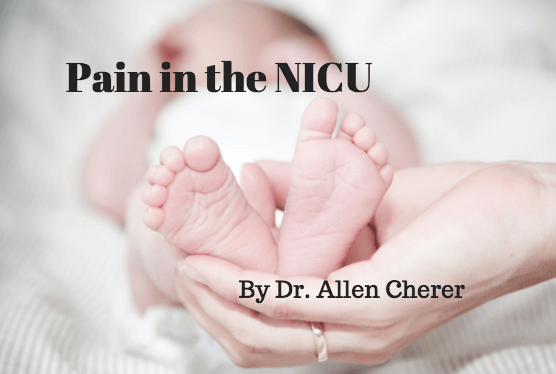
In addition, for the individual pregnant woman, a minimum of two lives is affected: her own and that of her unborn child. The prevalence of prenatally exposed newborns to one or more illicit drugs approximates 6%. Neonatal Abstinence Syndrome (NAS) refers to the withdrawal symptoms from physical dependence experienced by the newborn exposed during pregnancy generally to illicit drugs, prescribed drugs, or to those opioids employed in medication-assisted treatment of maternal opioid addiction.

Withdrawal symptoms can vary markedly in terms of time of onset and severity but typically manifest as tremulousness, agitation, sleeplessness, and poor feeding. NAS increased threefold from 2000-2009 and frequently requires prolonged newborn hospitalization. It has been reported that aggregate hospital charges for NAS increased from 732 million dollars to 1.5 billion dollars with approximately 80% attributed to state Medicaid programs in 2012. Clearly, NAS is a costly public health problem resulting in significant human suffering and expense.

Traditionally, infants who are known to be at risk for NAS have been monitored in the postpartum unit after birth for at least 96 hours and withdrawal symptoms scored based on the Finnegan Scale developed in the mid 1970’s. Typically, if the scores exceed certain values, the newborn is admitted to a Special Care Unit where pharmacologic treatment is frequently started. As withdrawal symptoms subside, dosing is gradually tapered and ultimately stopped. The newborn is observed off medication and monitored for recurrence of disabling withdrawal symptoms. The entire process can generally result in a prolonged Special Care Unit hospital stay of 2-10 weeks.

With the seemingly overnight explosion in the number of newborns demonstrating withdrawal symptoms in the early 2000’s, medical caregivers and hospitals were caught off-guard. On short notice, staff addiction education, medication and weaning protocols, general care policies, and hospital space allocation were required. After a number of years of concerted, collaborative work, much has been learned and achieved in improving the care of the substance-exposed infant.

Nevertheless, pharmacologic treatment continues to require prolonged hospital stays, often in costly Special Care Units. In addition, it effectively excludes full participation by the eventual sole primary caregivers, ideally the parents. It is with these disturbing issues in mind that it is refreshing to note the work and studies over the past several years to further optimize the care provided to infants with NAS and their families.

One of the earlier studies to suggest the therapeutic benefits of a different approach to caring for the drug-exposed infant was that of Abrahams et al. published in the Canadian Family Physician in 2007. During the same period of frenzy involving inpatient hospital transfers, guaranteeing interobserver scoring reliability, pharmacologic treatment protocols, and nursing care directives, the Canadian group with extensive previous experience in addiction medicine reported in a retrospective cohort study the benefits of a rooming-in policy whereby infants remained with their mothers as primary caretakers.

They noted that infants who roomed-in were less likely to require pharmacologic therapy for withdrawal and more likely to be discharged to mother’s care compared to infant’s who received standard nursery care. Subsequently, other retrospective cohort studies both in Europe and the United States demonstrated equally beneficial effects of rooming-in regarding decreased requirement for pharmacologic therapy and decreased duration of hospital stay.

Most recently, the results of a quality collaborative project from the Children’s Hospital at Dartmouth Hitchcock were described in the May, 2016 Pediatrics and demonstrated the beneficial effects of combined standardized protocols and family-centered care in the management of the drug-exposed infant. Over time, the project safely reduced the number of infants requiring pharmacologic therapy, average length of stay, and overall hospital costs.

Among others, key drivers to success were prenatal education of family caregivers including expressed expectation that they would provide meaningful rooming-in care, baby-centered NAS scoring including on demand feeding schedules, pharmacologic therapy when necessary with dosing adjustment based on overall infant condition rather than solely Finnegan score and determined by a consistent team, and an infant “snuggler” volunteer program to assist families when times required their absence.

Overall, the project demonstrated that despite many practical obstacles to providing high quality care for drug-exposed newborns and their families in the hospital setting, where there’s a will, there’s a way.

Antenatal Corticosteroid Use for Late Preterm Delivery

In 1972, Drs. Liggins and Howie published their landmark study demonstrating that antenatal corticosteroids administered to women 24-36 weeks of gestation reduced the incidence of respiratory distress syndrome and  neonatal mortality. Liggins had previously noted that lambs, treated with intrafetal  ACTH, cortisol, or dexamethasone, delivered prematurely, and sacrificed, demonstrated partially expanded  lungs.

Such alveolar stability was not typically noted until later in gestation. It suggested to Liggins that glucocorticoids might cause premature liberation of surfactant into the alveoli and served as the basis for his study. In the trial, the most significant difference in the incidence of respiratory distress syndrome among those treated vs. not treated with corticosteroids occurred in those gestations  less than 32 weeks.

Although those gestations treated between 32 and 37 weeks exhibited a decreased incidence of respiratory distress, the number did not reach statistical significance. Nevertheless, even at that time, Liggins postulated that mechanisms in addition to enhanced surfactant production and release might be responsible for the improved pulmonary function noted in more advanced gestations treated with antenatal corticosteroids.  

Interestingly, despite the findings of the initial study and similar results in multiple subsequent studies , the 1994 NIH Consensus report on the effect of corticosteroids for fetal maturation on perinatal outcomes found that only 20% of women who delivered newborns  501-1500 grams received the benefit of antenatal steroids. After a thorough review of available evidence, including  12 year neurodevelopmental follow up showing no adverse outcomes, the Consensus Panel felt  the benefits of antenatal administration of corticosteroids vastly outweigh the risks and all fetuses between 24 and 34 weeks gestation at risk of preterm delivery should be considered candidates for antenatal  treatment.

Only in those few pregnancies where corticosteroids would have an adverse effect on the mother or delivery was imminent  should steroid treatment be withheld. In addition, although Grade 1 evidence existed at the time to support the use of antenatal corticosteroids for gestations greater than 34 weeks, it was judged insufficient to recommend their use.

Since the Consensus statement, the use of antenatal corticosteroid use has become common and has resulted in considerable reduction in mortality and morbidity, as well as total health care costs. In addition, further neurodevelopmental follow up, including the original Auckland steroid trial participants, continues to demonstrate no adverse effects on psychological functioning and health-related quality of life. Other studies have demonstrated a decrease in overall respiratory disease in infants born beyond 34 weeks who had previously been exposed to antenatal corticosteroids when compared to unexposed infants born at similar gestations.

More than 300,000 pregnancies deliver in the late preterm period (34 0/7 – 36 6/7 weeks gestation) each year in the United States. Seventy per cent of Intensive Care Nursery admissions are late preterm newborns. Their increasing numbers and the broad range and severity of respiratory disorders with which they present beg for a re-evaluation of antenatal corticosteroid use in this range of gestations. This is especially appropriate with a better understanding of the multiple actions of corticosteroids as gestation approaches term.

A recent study, titled Antenatal Late Preterm Steroids (ALPS), a Randomized Trial to Reduce Neonatal Respiratory Morbidity, was published in The New England Journal of Medicine in April, 2016. The study enrolled over 2800 women with singleton pregnancies at high risk for late preterm delivery.

The participants were randomized to receive antenatal betamethasone by injection or a matching placebo. Greater than 80% of women in the trial delivered prior to 37 weeks gestation. The primary outcome was a neonatal composite of treatment in the first 72 hours (CPAP or High Flow Nasal Cannula for at least 2 hours, supplemental oxygen with fraction of inspired oxygen of at least 0.3 for at least 4 hours, mechanical ventilation, or ECMO) or stillbirth or neonatal death within 72 hours of birth.

The study found a significant decrease in neonatal respiratory complications in the group given the steroid treatment (11.6% vs. 14.4%). In addition, severe respiratory complications occurred significantly less frequently in the betamethasone group. The incidence of neonatal hypoglycemia  was increased in those treated with betamethasone (24% vs. 14.9%), but no other adverse neonatal outcomes were noted between the groups.

The study is authoritative due to its size, generalizability, and methodologic rigor. Although the issue of long term follow up cannot be specifically addressed, follow up studies of similar treatment in earlier gestations are reassuring. Late preterm births comprise a high risk group for hypoglycemia regardless of maternal antenatal steroid treatment and warrant vigilant monitoring during the newborn period.  In sum, the findings of the Antental Late Preterm Steroids study are consistent with other randomized controlled trials of antenatal corticosteroids administered at gestations less than 34 weeks.

Both the American College of Obstetrics and Gynecology with an endorsement by the American Academy of Pediatrics and the Society for Maternal-Fetal Medicine have addressed and published recommendations based on the study’s findings. Although the recommendations do not establish exclusive standards of care, the organizations approve the use of antenatal corticosteroids in certain defined late preterm pregnancies.  It is only with thoughtful application of the recommendations and further studies that the efficacy and safety of antenatal steroids in the late preterm pregnancy will be realized. It is a significant start.

Page 4 of 5

Powered by WordPress & Theme by Anders Norén