Dr. Allen Cherer is a neonatal care expert with over 30 years of medical accomplishments to his name.

Tag: Pediatrics Page 1 of 2

dr-allen-cherer-blood

A Closer Look at Neonatal Polycythemia

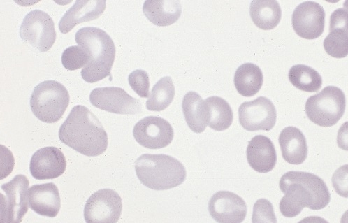
Polycythemia occurs when bone marrow is manufacturing too many red blood cells, thereby increasing the volume percentage of red blood cells in the blood. It can also be caused by a low plasma count in relation to blood cells, causing an imbalance. These excess cells, in turn, thicken the blood, which slows down the overall blood flow leading to more serious issues, such as blood clots. Polycythemia is essentially the opposite of anemia, which is when there are too few blood cells being produced. 

Neonatal is a term used to describe babies within the first 28 days of life. Neonatal polycythemia is diagnosed when a baby’s blood is composed of more than 65% red blood cells. This is a common problem with newborns, but that number is expected to decrease within a few hours if everything is normal. 

Some babies who are at a higher risk include those who are born small for their gestational age, babies who are post-term, infants of mothers with diabetes, twin-to-twin blood transfer in utero, low oxygen levels in fetal blood, and babies born with chromosomal abnormalities. 

It can be hard to diagnose this condition when babies are first born, because, aside from a high blood viscosity, babies might be asymptomatic before showing any metabolic changes due to this disorder. Samples should be taken from a largely free-flowing blood vessel in order to get an accurate hematocrit result. Signs that blood is affected include poor blood flow returning to a site after pressure is applied (peripheral perfusion), and blood having a ruddy, dusky appearance. There are also other clinical symptoms that include lethargy, irritability, tremors, seizures, lack of interest in feeding, hypoglycemia, rapid breathing (tachypnea), and bluish/grayish skin coloration (cyanosis).

There are several other conditions that share similar symptoms with neonatal polycythemia, which is why it is vital to rule them out before attempting any course of treatment. Some conditions that are similar include hypoglycemia, neological dysfunction, renal failure, or respiratory issues. Once a firm diagnosis is confirmed, there is a recommended course of action. 

Not all babies require treatment, but if there are signs of metabolic distress, the first priority is to lower the hematocrit by performing a partial exchange transfusion (PET). Either a saline solution may be used or a 5% protein solution. Saline is the preferred material because it won’t risk infection and has a better price point. It is strongly advised to avoid fresh frozen plasma because studies have shown a correlation between its use and necrotizing enterocolitis.

dr-allen-cherer-babyhead

A Closer Look at Neonatal Macrosomia

Neonatal macrosomia refers to babies weighing more than eight pounds and 13 ounces at birth. Approximately nine percent of infants are born with the condition. The larger the baby, the greater the risks to the mother and the infant. There are a variety of causes and risk factorsYour text to link… that lead to overweight newborns. Some causes are preventable.

Causes and Risks

  • Diabetes-Expectant mothers may have been diagnosed with diabetes before becoming pregnant. Others develop gestational diabetes during pregnancy. Blood sugars must be monitored and controlled otherwise, the infant develops with a larger amount of body fat.
  • Previous history-Women who have given birth to overly large infants in the past have a greater risk of having large babies in the future.
  • Obesity-There is a greater chance of having a baby with neonatal macrosomia if the mother is obese. Gaining too much weight during pregnancy also increases the risk.
  • Male infants-Neonatal macrosomia occurs more often in boy babies.
  • Overdue pregnancies-Pregnancies that extend two or more weeks beyond the estimated due date increase the chance that the infant will be overly large.
  • Mother’s age-Pregnant women over the age of 35 are more likely to have abnormally large babies.

Maternal Complications

  • Difficult labor-When an infant is too large, there is a likelihood that the baby becomes stuck in the birth canal, which may necessitate a C-section delivery.
  • Internal injuries-During the birthing process, the mother may suffer laceration or tearing of the vaginal tissues and perineal muscles.
  • Hemorrhaging-Internal injuries combined with the uterus’ inability to contract properly may lead to severe bleeding.
  • Uterine damage-Women who previously gave birth via C-section or had gynecological surgery have an increased risk of suffering from a uterine rupture.

Infant Complications

  • Hypoglycemia-Babies born with neonatal macrosomia have an increased risk of suffering from abnormally low blood sugars.
  • Obesity-Overly large infants are at a greater risk of becoming obese during childhood.
  • Metabolic syndrome-Neonatal macrosomia infants are likely to have metabolic syndrome. The condition is associated with hypertension, hyperglycemia, elevated cholesterol and excess body fat.

Prevention

Women must maintain a healthy weight before during and after pregnancy. While pregnant women should not gain ore than 35 pounds. Women diagnosed with diabetes must have their blood sugar continually monitored and controlled.

dr-allen-cherer-baby

Diagnosing and Addressing Neonatal Apnea

Apnea is a term defined as the cessation of breathing for longer than 10 to 15 seconds. While this can happen at any age, it typically affects infants aged two to four weeks until six months.

It is particularly seen in premature infants born around 28 weeks due to their underdeveloped respiratory systems. It happens when the brain and spinal cord do not mature, obstructing breathing

When apnea develops, it can have many causes. The most common links found in neonatal apnea are an infection, neurological, cardiovascular, pulmonary, metabolic, temperature regulation and maternal drug use.

Types of Apnea

There are three main types of apnea. These are central, obstructive and mixed apnea.

Central apnea is when there is no signal of breathing transmitted to the respiratory muscles, causing the system to not respond due to immature development.

Obstructive apnea is when there is a brief pause of airflow in the pharynx where the muscles are too weak to help the infant breathe properly.

Mixed apnea is a combination of the two.

Treating Neonatal Apnea

To manage apnea in infants born before 34 weeks gestation, it is important for professionals in the neonatal intensive care unit to monitor breathing and development. Underlying causes will also have to be determined and close monitoring is imperative. Health professionals will check to see if there is a link to bradycardia and hypoxia.

Bradycardia is a heart rate that is too slow for normal functionality. Hypoxia is when there is an oxygen distribution deprivation. These two conditions are often linked to cases of apnea in infancy.

Management varies between infants and will depend on a series of factors. Medicines will be administered depending on the severity and cause of the issue.

Untreated apnea can cause unwanted effects to the overall wellbeing of the child. These effects can be a failure to thrive or decrease in intellect. Certain types of apnea can also result in death.

Having a wide group of trained health professionals can assist in the monitoring and betterment of neonates. Once proper diagnosis and treatment are implemented, the infant can be treated accordingly until the risks decline and their health improves.

dr-allen-cherer-chest

Exploring Neonatal Sepsis

Neonatal Sepsis is a blood infection that infants may develop before reaching 90 days of age. Babies can also develop early-onset and late-onset sepsis.

Causes?

A bacteria named Eschericia coli (E coli) and Listeria can cause infants to develop sepsis. A specific streptococcus strain (Group B streptococcus or GBS) can also make an infant ill. If the baby’s mother contracts herpes simplex virus (HSV), this can also lead to neonatal sepsis.

An early-onset case usually develops 24 to 48 hours after the baby’s birth, usually by being exposed during birth. 

Contributors to early-onset sepsis:

  • Preterm delivery
  • GBS colonization during mother’s pregnancy
  • Placental tissues and amniotic fluid become infected (chorioamniontitis)
  • Early rupture of membranes (more than 18 hours)

Late-onset sepsis risks:

  • Extended hospitalization for infant
  • Keeping a catheter in baby’s blood vessel for an extended time

Symptoms?

  • Breathing problems
  • Changes in body temperature
  • Decreased bowel movements or diarrhea
  • Reduced movements
  • Low blood sugar
  • Reduced suckling
  • Heart rate is fast or slow
  • Seizures
  • Vomiting
  • Swollen abdomen
  • Jaundice (yellow skin and whites of eyes)

Diagnostic Tests?

Pediatricians perform the following diagnostic tests:

  • C-reactive protein
  • Blood culture
  • Complete blood count (CBC)
  • Lumbar puncture
  • Urine, skin or stool cultures to search for herpes virus
  • Chest X-ray (if baby has difficulty breathing)
  • Urine cultures

Treatments?

Even if the newborn is symptom-free, they will receive intravenous antibiotics. Babies younger than 4 weeks with fever or other symptoms receive IV antibiotics immediately.

The baby stays on antibiotics for three weeks if bacteria is in the spinal fluid or blood. This is shorter if no bacteria is present.

Acyclovir (antiviral medication) is given for HSV-caused infections.

If the baby has already gone home, it will be re-admitted to the hospital for treatment.

Outlook?

The infant may recover completely and show no evidence of any other problems. Neonatal sepsis can lead to infant death. The sooner treatment starts, the better the prognosis.

Potential Complications?

  • Disability after illness
  • Death

Prevention?

Pregnant mothers should receive preventive antibiotics if they have these illnesses:

  • Group B strep colonization
  • Chorioamnionitis
  • Has already had a baby with bacterial sepsis
  • This condition is preventable. Babies should be delivered 12 to 24 hours after water breaks.

Other Names?

Other names include:

  • Neonatal septicemia
  • Sepsis – infant
  • Sepsis neonatorium
dr-allen-cherer-echogenic

Echogenic Bowel: An Overview

Though it is not an incredibly common diagnosis, there are still thousands of fetuses diagnosed with an echogenic bowel each year. Roughly 1.8 percent of all fetuses have echogenic bowel, and their parents often wonder what exactly this term means. Understanding all the details about echogenic bowel will help parents discover how to handle this condition.

What Is Echogenic Bowel?

This term is simply used to refer to a fetal bowel that is abnormally bright when viewed on an ultrasound. Ultrasounds are always in shades of black, grey, and white, with denser tissue being bright white and fluid being black. Usually, the bowels of the fetus tend to be a dark grey on an ultrasound because they are softened tissue, but if a fetus has echogenic bowel, their bowels may look as bright as thick bones like the pelvic bone.

What Does It Mean to Have Echogenic Bowel?

When parents first hear this word, they often start to wonder if it is a serious problem. The reality is that it is not always a sign of a health condition. 0.5 percent of all perfectly healthy fetuses have an echogenic bowel, and it can just occur due to various fluctuations in growth. However, echogenic bowel can be a cause for concern because it is more common in babies born with Down syndrome or cystic fibrosis. It can also be a sign that the fetus is suffering from an intestinal obstruction or an infection like cytomegalovirus or toxoplasmosis.

How Is Echogenic Bowel Treated?

The method for addressing echogenic bowel typically depends on the severity. Low grade echogenic bowels which are less dense than bone are normally harmless, so doctors tend to take a “wait and see” approach. They normally recommend taking a more detailed ultrasound in a few weeks to check up on the growth of the fetus. However, for more severely echogenic bowels, doctors may recommend a maternal serum screening, blood tests, or an amniocentesis to determine if the fetus has Down syndrome, infections, or cystic fibrosis. To make sure that the pregnancy continues safely, doctors will usually do regular fetal monitoring to make sure the fetus is growing properly following a display of echogenic bowel.

dr-allen-cherer-dna

DNA Sequencing Could Change How We Look at Genetic Neonatal Diseases

DNA sequencing is one of the most promising new technologies in terms of identifying the risk of disease, but it might not quite be ready for market. But regardless of concerns that DNA sequencing isn’t yet a safe screening method for newborn infants, chances are very strong that it will become a regular toolkit in preventative medicine sooner rather than later.

Routine blood tests are already part of standard procedure for infants born in the United States, and these tests can provide some substantive insight into potential future risks. But while a routine blood test can help identify dozens of different genetic conditions, that’s just scratching the surface of what can be accomplished with DNA sequencing. A study published in the American Journal of Human Genetics conducted DNA sequencing on 159 babies and found that 9% displayed anomalies that could predict genetic diseases that could appear in childhood. These include congenital heart disease and hearing loss.

But how much of an effect this testing could have on the health of infants is still an open question. Even co-author Alan Beggs questions how much substantive and actionable intelligence will arise from these genetic markers, at least for now. Nine percent is a low number, and many of these issues can be uncovered with the existing blood testing. Then there’s the fact that many of these genetic markers are not that well understood yet, and it can be difficult to understand how high of a risk such a genetic marker would actually pose.

Finally, there are a number of ethical and practical questions to consider. It can be hard to unpack issues of consent when dealing with the very genetic makeup of a child, and the rules behind the sharing of personal data, even as a means to better understand the map of human DNA, is still something like the Wild West. Finally, there are questions of how accessible this technology is and the costs associated for both medical providers and patients.

The running consensus now seems to be that DNA sequencing may be a beneficial choice in specific instances where parents are concerned about severe genetic disorders, but it’s not quite ready for primetime. As the technology and research continues to develop, it will likely become standard practice as a complement rather than a replacement for standard and accepted blood tests.

dr-allen-cherer-hepb

Fine-tuning the Elimination of Perinatal Hepatitis B Infection

Hepatitis B virus (HBV) infection  is a serious illness in the newborn and young infant.  The virus,  first discovered in the mid-1960s, is transmitted through percutaneous (i.e., puncture through skin) or mucosal (i.e., direct contact with mucous membranes) exposure to infectious blood or body fluids. The  virus is highly infectious, can be transmitted in the absence of visible blood, and remains viable on environmental surfaces for at least seven days.  Once the virus enters the body, it is transported to the liver where it replicates.  Although one generally thinks of the acute illness as a self-limited one in the adult with characteristic signs and symptoms, HBV infection in the infant is almost exclusively asymptomatic and hence, unrecognized. The devastating aspect of the infection is that the infant and young child frequently fail to clear the virus, and the illness becomes chronic. As many as 80-90% of infected infants progress to chronic infection, and chronically infected persons as adults are at increased risk of cirrhosis, hepatocellular carcinoma, and liver failure with approximately 25% dying from these serious complications.

Before 1982, an estimated 200,000-300,000 persons in the U.S. alone were infected with HBV annually, including approximately 20,000 infants. No effective pre-exposure prophylaxis existed, and only post-exposure prophylaxis in the form of hepatitis B immune globulin (HBIG) was available. However, the first hepatitis B vaccine was approved in the United States in 1981 and proved to be a real game changer. The availability of the vaccine set the stage for remarkable progress in the elimination of HBV infection among all age groups. With the advent of an effective vaccine, incurable hepatitis B infection had become preventable. The vaccine saves lives!

It is in this setting of disease prevention  through widespread vaccination that an evolving strategy to eliminate perinatal hepatitis B infection was initiated over 30 years ago. Early epidemiological studies had demonstrated that a major contributor to perinatal HBV infection is mother-to-child transmission  (MTCT) at the time of delivery. In utero infection is felt to account for less than 2% of infections. The risk of transmitting the virus was estimated to be 20-80% depending on the activity of the maternal infection. Initial attempts in the early 1980s to limit vertical HBV transmission were risk-based and aimed at identifying those pregnant women considered infectious by virtue of the serum marker, HBsAg. With reliable identification of mothers and expeditious treatment of their newborns with hepatitis B vaccine and HBIG, HBV infection could be prevented. However, it became clear within several years that such screening was inadequate with as many as 35-65% of HBsAg-positive women being missed. Consequently in 1988, the Advisory Committee on Immunization Practices (ACIP) of the Centers for Disease Control and Prevention recommended universal testing of all women early in each pregnancy such that at risk babies would receive appropriate post-exposure HBV prophylaxis. Throughout the 1990s, efforts were intensified to eliminate all HBV-related  illness through widespread vaccination of children, adolescents, and at-risk adults. Studies showed that receipt of the 3-dose hepatitis B vaccine series produced a protective antibody response in approximately 98% of healthy infants. During 1990-2004, the incidence of acute hepatitis B in the U.S. declined by 75%. The greatest decline (94%) occurred among children and adolescents, most likely due to increasing hepatitis B vaccine coverage. As of 2004, over 92% of children less than 3 years of age had been fully vaccinated with the complete series.

Coupled with the remarkable success of the hepatitis B elimination strategy is the knowledge that the task is not complete. As a  response to the persistence of perinatal HBV infection  and aware that errors in testing as well as in communication of results may occur, ACIP has recommended a change in the administration of the initial hepatitis B vaccine dose over time. Initially, the first dose could be administered to an infant born to a HBsAg-negative mother any time from birth to 2 months of age.  Subsequently the initial dose became the “birth dose” with the recommendation that it be given prior to discharge, and in 2017, the initial dose was to be administered within 24 hrs of birth. The previous  permissive language that allowed the dose to be delayed “on a case-by –case basis and only in rare circumstances” was omitted. Based on the fact that the vaccine alone is 75% effective in preventing MTCT, these changes reflect reality and provide basic protection. Then too, the emerging concept that maternal viral load (HBV DNA) plays a significant role in risk of MTCT now plays a prominent role in management.  Testing pregnant HBsAg-positive women for HBV DNA is now recommended to guide the use of antiviral therapy during the third trimester for the purpose of preventing perinatal HBV transmission.

Congenital Hypothyroidism and Newborn Screening

Congenital Hypothyroidism and Newborn Screening

Newborn screening for Congenital Hypothyroidism (CH) is a major public health achievement. Thyroid hormone is essential for the maturation of brain function and somatic growth, and its deficiency early in life can lead to mental retardation. For the fetus, maternal thyroid status is important during the first half of gestation; thereafter, the fetus’  hypothalamus-pituitary-thyroid axis is functional in the normal situation. For the hypothyroid newborn, it is well documented that provision of thyroid hormone is critical during the first weeks of life to avoid severe intellectual impairment. Notably, congenital hypothyroidism is considered one of the most common preventable causes  of mental retardation.

Studies showed that affected newborns were rarely identified during the first months of life and were often missed until 1-3 years of age. Congenital hypothyroidism  was found to be  an ideal candidate with the introduction of dried blood newborn screening by Dussault in Canada. With the development of increasingly sensitive assays to measure thyroid hormone (T4) and thyroid stimulating hormone (TSH) using a dried blood spot (DBS), newborn screening programs have developed throughout much of the world. In the 1980s, the incidence of CH in the United States was estimated to be 1:3000-1:4000. More recently, screening programs have reported an increased incidence of 1:1400-1:2800, most probably due to changes in screening strategies and the identification of milder cases.

Typically, newborn screening requires a heel stick blood specimen obtained at 48-72 hrs of life prior to an infant’s discharge from the hospital. Most current assays measure TSH alone as an indicator of thyroid function.  Results above established cutoff levels generally signify thyroid gland dysfunction and indicate further testing. Although most helpful in early identification of term newborns with anatomic or functional thyroid gland abnormalities, the screening does miss a percentage of newborns, for example those with central hypothyroidism due to hypothalamic-pituitary failure and the increasingly larger group of preterm  infants with congenital hypothyroidism who demonstrate delayed elevations in TSH. Numerous questions remain regarding the optimal timing of follow up laboratory studies and even treatment of certain types of newborn thyroid dysfunction.Nevertheless, newborn screening has proved invaluable for the great number of affected newborns.

The American Academy of Pediatrics recommends the measurement of TSH in all newborns with the goal that all infants with CH be identified by 2 weeks of age and that effective treatment with thyroid hormone replacement be started such that serum TSH levels less than 5 mIU/L be achieved within 4 weeks of diagnosis. Unfortunately, despite the significant successes following early identification and treatment of newborns with CH, obstacles persist in reaching the Academy’s goals. Screening programs continue to be plagued with the practical problems of screening all newborns, particularly those discharged home early who are lost to recall or lost to follow up altogether.  In addition, dried blood specimens are collected or processed improperly. Delays occur with recall of infants with abnormal results and with appropriate referrals for definitive treatment and management. A recent study conducted in Utah and reported at the 86th Annual Meeting of the American Thyroid Association highlights some of the problems which currently exist. After reviewing the TSH assays of 4394 children under 2 years of age, 48% of initial samples with elevated  levels (>20 mIU/L) were obtained after  the first 2 weeks of life, 15% of the initial abnormal TSH assays were not retested, and only 34% of those infants with initial elevated TSH assays achieved the goal of TSH < 5 mIU/L within 28 days of the initial assay.

The final message is that it is not enough to rely on the known efficacy of newborn screening for congenital hypothyroidism, but greater vigilance must be exercised to maximize its benefits in the lives of children.

Ehrenkranz J, Butler A, Snow G, Bach P. oral Abstract 19. The Diagnosis and Treatment of Congenital Hypothyroidism in Utah 2006-2015. Presented at: American Thyroid Association Annual Meeting; September 21-25, 2016; Denver, Colorado

Noting the Extraordinary Success of Hib Vaccination

August is observed as National Immunization Awareness Month and is a time to highlight the extreme importance and value of vaccination for people of all ages. Vaccination serves as one of the best ways to protect infants, children, and adolescents from sixteen potentially harmful, and even deadly, diseases. Although it is common to think of the vaccines against measles, pertussis, and polio, an astonishingly important vaccine since the end of the 20th century has targeted the bacteria, Haemophilus influenzae type b (Hib).

Haemophilus influenzae is a small, pleomorphic, gram negative coccobacillus. Some strains of H. influenzae possess a polysaccharide capsule, and these strains are serotyped into six different types (a-f) based on their biochemically different capsules.

The H. influenzae strains with no capsule are termed nonencapsulated H. influenzae or nontypable H. influenzae (NTHi). H. influenzae type b is the most virulent, with its polysaccharide capsule being the main factor. Antibody to the capsule is the primary contributor to serum bactericidal activity, and increasing levels of antibody are associated with decreasing risk of invasive H. influenzae disease.

H. influenzae type b most commonly causes pneumonia, bacteremia, meningitis, epiglottitis, and cellulitis. Non-type b encapsulated forms present in a similar manner to type b infections, while non typable strains more commonly cause infections of the respiratory tract, such as pneumonia, otitis media, sinusitis, and conjunctivitis.

Generally, the mode of transmission is person to person by inhalation of respiratory tract droplets or by direct contact with respiratory tract secretions. Pharyngeal colonization by H. influenzae is relatively common, especially with nontypable and non-type b capsular strains.
Before effective Hib conjugate vaccines for infants older than 2 months were available in 1990, Haemophilus influenzae type b was the leading cause of invasive bacterial disease among children in the United States.

One in 200 children developed invasive Hib disease by 5 years of age; approximately 60% of these children had meningitis and 3-6% died from the disease. Of the Hib meningitis survivors, many exhibited permanent sequelae ranging from mild hearing loss to mental retardation.

Sadly, I recall as a Pediatric resident admitting to the hospital at least one infant with H. influenzae type b meningitis almost every night when on call.Remarkably, since the introduction of Hib conjugate vaccines in the United States, the incidence of invasive Hib disease has decreased a stunning 99% to fewer than 1 case/100,000 children younger than 5 years of age, and in 2012, only 30 cases of invasive type b disease were reported in children under 5 years old.

Truly, it has been an amazing accomplishment. Nevertheless, the risk for invasive Hib disease persists among unimmunized and underimmunized children, highlighting the importance of full vaccination with the 2 or 3 injection (depending on the product) series between 2 and 6 months old and a single booster dose given between 12 and 15 months of age.

Certain additional doses may be indicated over 5 years of age depending on medical conditions, such as anatomic or functional asplenia, hematopoietic stem cell transplantation, or HIV infection. The Hib vaccine is very safe. The most common side effects are usually mild and consist of fever and rednesss, swelling, or warmth at the injection site. As with all current vaccines, significant advances and improvement in public health have been witnessed. It is incumbent upon each of us to maintain that success.

Providing Care for Drug-exposed Newborns: Time for the Next Step

During the years 1999-2013, the amount of prescription opioids dispensed in the United States nearly quadrupled, and since 2000, it is estimated that opioid use during pregnancy has tripled. Notably, the tragic consequences of the extreme availability of such drugs include abuse, physical dependence, and increasingly, death through inadvertent overdose.

newborn-boy-sleepingIn addition, for the individual pregnant woman, a minimum of two lives is affected: her own and that of her unborn child. The prevalence of prenatally exposed newborns to one or more illicit drugs approximates 6%. Neonatal Abstinence Syndrome (NAS) refers to the withdrawal symptoms from physical dependence experienced by the newborn exposed during pregnancy generally to illicit drugs, prescribed drugs, or to those opioids employed in medication-assisted treatment of maternal opioid addiction.

Withdrawal symptoms can vary markedly in terms of time of onset and severity but typically manifest as tremulousness, agitation, sleeplessness, and poor feeding. NAS increased threefold from 2000-2009 and frequently requires prolonged newborn hospitalization. It has been reported that aggregate hospital charges for NAS increased from 732 million dollars to 1.5 billion dollars with approximately 80% attributed to state Medicaid programs in 2012. Clearly, NAS is a costly public health problem resulting in significant human suffering and expense.

Traditionally, infants who are known to be at risk for NAS have been monitored in the postpartum unit after birth for at least 96 hours and withdrawal symptoms scored based on the Finnegan Scale developed in the mid 1970’s. Typically, if the scores exceed certain values, the newborn is admitted to a Special Care Unit where pharmacologic treatment is frequently started. As withdrawal symptoms subside, dosing is gradually tapered and ultimately stopped. The newborn is observed off medication and monitored for recurrence of disabling withdrawal symptoms. The entire process can generally result in a prolonged Special Care Unit hospital stay of 2-10 weeks.

With the seemingly overnight explosion in the number of newborns demonstrating withdrawal symptoms in the early 2000’s, medical caregivers and hospitals were caught off-guard. On short notice, staff addiction education, medication and weaning protocols, general care policies, and hospital space allocation were required. After a number of years of concerted, collaborative work, much has been learned and achieved in improving the care of the substance-exposed infant.

Nevertheless, pharmacologic treatment continues to require prolonged hospital stays, often in costly Special Care Units. In addition, it effectively excludes full participation by the eventual sole primary caregivers, ideally the parents. It is with these disturbing issues in mind that it is refreshing to note the work and studies over the past several years to further optimize the care provided to infants with NAS and their families.

One of the earlier studies to suggest the therapeutic benefits of a different approach to caring for the drug-exposed infant was that of Abrahams et al. published in the Canadian Family Physician in 2007. During the same period of frenzy involving inpatient hospital transfers, guaranteeing interobserver scoring reliability, pharmacologic treatment protocols, and nursing care directives, the Canadian group with extensive previous experience in addiction medicine reported in a retrospective cohort study the benefits of a rooming-in policy whereby infants remained with their mothers as primary caretakers.

They noted that infants who roomed-in were less likely to require pharmacologic therapy for withdrawal and more likely to be discharged to mother’s care compared to infant’s who received standard nursery care. Subsequently, other retrospective cohort studies both in Europe and the United States demonstrated equally beneficial effects of rooming-in regarding decreased requirement for pharmacologic therapy and decreased duration of hospital stay.

Most recently, the results of a quality collaborative project from the Children’s Hospital at Dartmouth Hitchcock were described in the May, 2016 Pediatrics and demonstrated the beneficial effects of combined standardized protocols and family-centered care in the management of the drug-exposed infant. Over time, the project safely reduced the number of infants requiring pharmacologic therapy, average length of stay, and overall hospital costs.

Among others, key drivers to success were prenatal education of family caregivers including expressed expectation that they would provide meaningful rooming-in care, baby-centered NAS scoring including on demand feeding schedules, pharmacologic therapy when necessary with dosing adjustment based on overall infant condition rather than solely Finnegan score and determined by a consistent team, and an infant “snuggler” volunteer program to assist families when times required their absence.

Overall, the project demonstrated that despite many practical obstacles to providing high quality care for drug-exposed newborns and their families in the hospital setting, where there’s a will, there’s a way.

Page 1 of 2

Powered by WordPress & Theme by Anders Norén