Dr. Allen Cherer is a neonatal care expert with over 30 years of medical accomplishments to his name.

Tag: Neonatal Care Page 1 of 2

Screen Shot 2020 07 30 At 11.48.40 Am

Neonatal Septal Defects: An Overview

Ventricular septal defects, or VSD, refers to an opening in the wall that separates the lower ventricles of the heart. The condition occurs naturally in neonates at a rate of one out of every 250 to 330 births. Normally, the hole closes before the infant is born, which prevents oxygenated blood from combining with unoxygenated blood.


Under normal circumstances, blood enters the right side of the heart and continues into the lungs to receive oxygen. The blood then travels to the left side of the heart and is pumped through the body. But, when VSD occurs, more blood enters the lungs than normal, which stresses the heart and the lungs.


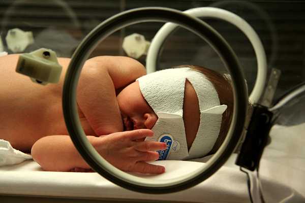
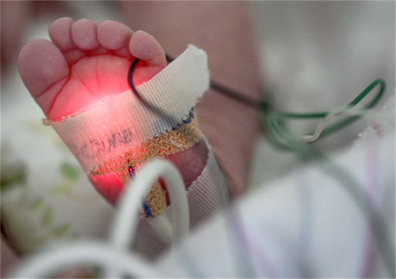
If the hole is small, physicians might hear a murmur when listening to the heart using a stethoscope. Otherwise, the child exhibits no symptoms. The opening is not large enough to add stress. However, if the hole is large, the infant breathes faster and harder than normal secondary to the stress on the heart and lungs. They may also exhibit difficulties when suckling and gasp for breath. Symptoms may occur shortly after the birth of the child. Or, the signs may not appear until weeks later when the lungs become hypertensive. If the child does not receive medical intervention, the lungs and blood vessels may endure irreversible damage.


Small hole defects commonly close without intervention. However, if the defect is deemed to be large, surgical repair is required. The procedure used depends on the size of the defect. Some are easily corrected in a cath lab. If the hole is not extremely large, surgeons may simply sew the detector closed. Other options include surgically applying a fabric or tissue patch over the hole. The patch is later naturally covered by normal tissue that lines the heart.


Banding the pulmonary artery is another option, which reduces the amount of blood that flows into the lungs. The scheduled surgery takes place anytime from early infancy into later childhood depending on the severity of the condition and accompanying symptoms.


Once the defect is medically corrected, the infant or child may resume a normal life. A pediatric cardiologist may advise that the child undergo periodic evaluations to ensure the ongoing health and detect possible complications. In rare cases, a heart valve may develop a leak once the child is older, which also requires intervention.

dr-allen-cherer-pregnancy

Top Pregnancy Myths: 2020

Some of the information expectant mothers receive is often based on myths or old wives’ tales. Dispelling the myths may bring comfort and reassurance in addition to ensuring the health of the expectant mother and growing infant.

You’re Eating for Two

For decades, women were encouraged to substantially increase their dietary intake in order to ensure they were consuming enough nutrients for the growing infant. However, overeating leads to obesity, which leaves the mother and baby at risk. Being overweight increases the chances of developing gestational diabetes or hypertension. The excess weight also stresses the cardiovascular system. Health care providers suggest that increasing daily calorie intake by a mere 200 to 300 calories is more than sufficient to ensure a healthy pregnancy.

Belly Size and Shape Reveals Gender

Physicians rebuke the belief that external appearance correlates with the baby’s gender. Some women carry the baby high while others carry it lower. However, the difference is often equated with genetics and physical characteristics and not infant gender.

Moisturizing Prevents Stretch Marks

Cocoa butter has long been touted as being one of the solutions to prevent stretch marks. While moisturizing preparations are good for the skin, they do not prevent the physiological effects that a growing infant causes on external skin. Women develop varying degrees of marks depending on genetics and the extent that the abdomen needs to stretch to accommodate the infant.

Stay Away from Cats

There is no reason why expectant mothers cannot have and care for a feline companion. The danger lies in changing the litter box. Feline waste products commonly contain a parasite that has the potential for causing toxoplasmosis. While the mother may or may not experience flu-like symptoms, the illness has the potential of becoming serious in infants. Best to leave litter box duties to someone else. The disease can also be contracted by eating undercooked meat or unwashed fruits and vegetables.

Jaundice Phototherapy

A Closer Look at Neonatal Jaundice

Neonatal jaundice is caused by unconjugated bilirubin in a newborn’s body. It’s presently the most common condition that requires medical attention or hospital readmission for newborns. This condition can have dire consequences, so early detection and treatment is crucial.

Symptoms Of Neonatal Jaundice

It’s common practice for hospitals to check newborns for jaundice before release,however parents should be aware of what to look for. Some symptoms of jaundice in infants include:

  • Yellow sclera (white part of the eyes)
  • Yellow skin, especially the abdomen and limbs
  • Baby is hard to awaken or lethargic
  • Baby is not gaining weight

If these symptoms appear after a child has been sent home from the hospital, it may be advisable to bring them back in for medical treatment.

Causes Of Neonatal Jaundice

Jaundice in infants can often be caused by the liver being underdeveloped. This leads to an inability for the liver to properly break down the bilirubin in their bodies. Besides immature livers, some other causes of this condition include:

  • Blood infection (sepsis)
  • Viral or bacterial infection
  • Malfunction of the liver
  • Deficient enzymes
  • Internal bleeding
  • Abnormal red blood cells

If symptoms of jaundice appear, the child will need to be checked for underlying causes to form a treatment plan.

Factors That Increase Risk Of Neonatal Jaundice

There are some factors that make jaundice more likely in infants. These factors include:

  • Bruising during birth
  • Mother and child’s blood types are different
  • Birth before 38 weeks
  • Breastfeeding difficulties

If a child has these risk factors, their parents and medical team will need to monitor them.

Treatment Of Neonatal Jaundice

To avoid acute or chronic complications, jaundice needs early treatment. Some ways it can be treated include:

  • Light therapy
  • Intravenous immunoglobulin
  • Exchange transfusion

These methods focus on diluting pathogenic antibodies in the child’s blood that cause jaundice or, in the case of light therapy, help the child’s body to more easily break down and excrete the excess bilirubin.


dr-allen-cherer-chest

Exploring Neonatal Sepsis

Neonatal Sepsis is a blood infection that infants may develop before reaching 90 days of age. Babies can also develop early-onset and late-onset sepsis.

Causes?

A bacteria named Eschericia coli (E coli) and Listeria can cause infants to develop sepsis. A specific streptococcus strain (Group B streptococcus or GBS) can also make an infant ill. If the baby’s mother contracts herpes simplex virus (HSV), this can also lead to neonatal sepsis.

An early-onset case usually develops 24 to 48 hours after the baby’s birth, usually by being exposed during birth. 

Contributors to early-onset sepsis:

  • Preterm delivery
  • GBS colonization during mother’s pregnancy
  • Placental tissues and amniotic fluid become infected (chorioamniontitis)
  • Early rupture of membranes (more than 18 hours)

Late-onset sepsis risks:

  • Extended hospitalization for infant
  • Keeping a catheter in baby’s blood vessel for an extended time

Symptoms?

  • Breathing problems
  • Changes in body temperature
  • Decreased bowel movements or diarrhea
  • Reduced movements
  • Low blood sugar
  • Reduced suckling
  • Heart rate is fast or slow
  • Seizures
  • Vomiting
  • Swollen abdomen
  • Jaundice (yellow skin and whites of eyes)

Diagnostic Tests?

Pediatricians perform the following diagnostic tests:

  • C-reactive protein
  • Blood culture
  • Complete blood count (CBC)
  • Lumbar puncture
  • Urine, skin or stool cultures to search for herpes virus
  • Chest X-ray (if baby has difficulty breathing)
  • Urine cultures

Treatments?

Even if the newborn is symptom-free, they will receive intravenous antibiotics. Babies younger than 4 weeks with fever or other symptoms receive IV antibiotics immediately.

The baby stays on antibiotics for three weeks if bacteria is in the spinal fluid or blood. This is shorter if no bacteria is present.

Acyclovir (antiviral medication) is given for HSV-caused infections.

If the baby has already gone home, it will be re-admitted to the hospital for treatment.

Outlook?

The infant may recover completely and show no evidence of any other problems. Neonatal sepsis can lead to infant death. The sooner treatment starts, the better the prognosis.

Potential Complications?

  • Disability after illness
  • Death

Prevention?

Pregnant mothers should receive preventive antibiotics if they have these illnesses:

  • Group B strep colonization
  • Chorioamnionitis
  • Has already had a baby with bacterial sepsis
  • This condition is preventable. Babies should be delivered 12 to 24 hours after water breaks.

Other Names?

Other names include:

  • Neonatal septicemia
  • Sepsis – infant
  • Sepsis neonatorium
dr-allen-cherer-neonatology

Neonatology: a Brief History

Physicians and scientists began recognizing that premature or ill newborns required specialized care in the 1700s. However, it would be another century before a physician would take the first steps toward improving neonatal health. In the coming years, advancements in science and technology steadily enhanced the chances that preterm infants survived.

19th Century 

French obstetrician Etienne Stephane Tarnier recognized that premature infants were unable to maintain their body temperature. The physician invented the first incubator using a wooden box with a glass lid. The heat was provided by a hot water bottle. As a result, infant mortality decreased by 28 percent.

Pierre-Constant Budin trained under Dr. Tarnier and became a pioneer in neonatal nutrition during the late 1800s. Dr. Budin was aware of the risks of feeding newborns cow’s milk due to pathogens. He encouraged his new mothers to breastfeed. He was also responsible for introducing tube feeding for preemies who were unable to feed naturally.

By the early 1900s, Martin Couney, one of Dr. Budin’s students, improved upon Tarnier’s incubator design. However, the medical community was not accepting and the devices were not used in hospitals. In order to gain attention for the need, Dr. Couney began treating infants free of charge and demonstrated his invention at expositions and fairs.

20th Century 

For the most part, premature or ailing infants were not provided medical care. It was not until after World War II that the medical community recognized the need to offer specialized care. During this era, hospitals began developing “Special Care Baby Units” that eventually evolved into NICUs. Along with providing sufficient warmth, the units ensured that the infants received oxygen. There was also increasing awareness of an infant’s susceptibility to infection, which led to stringent hand washing.

Formulas for premature infants were introduced during this time. The formulas contained increased levels of calcium, phosphorus, sodium and protein. However, the high protein levels soon created a number of problems. As such, whey proteins were used.

Beginning in the 1960s, laboratory tests and values were established to monitor infant health. Physicians created a way to evaluate blood gases, bilirubin levels and liver function along with checking electrolytes, blood sugar and oxygen levels.

Advancements in knowledge and technology meant that infants born after 23 weeks of gestation had a survival rate of 33 percent. Infants born after 24 weeks had a survival rate of 66 percent. The survival rates continue growing each year.

dr-allen-cherer-dna

DNA Sequencing Could Change How We Look at Genetic Neonatal Diseases

DNA sequencing is one of the most promising new technologies in terms of identifying the risk of disease, but it might not quite be ready for market. But regardless of concerns that DNA sequencing isn’t yet a safe screening method for newborn infants, chances are very strong that it will become a regular toolkit in preventative medicine sooner rather than later.

Routine blood tests are already part of standard procedure for infants born in the United States, and these tests can provide some substantive insight into potential future risks. But while a routine blood test can help identify dozens of different genetic conditions, that’s just scratching the surface of what can be accomplished with DNA sequencing. A study published in the American Journal of Human Genetics conducted DNA sequencing on 159 babies and found that 9% displayed anomalies that could predict genetic diseases that could appear in childhood. These include congenital heart disease and hearing loss.

But how much of an effect this testing could have on the health of infants is still an open question. Even co-author Alan Beggs questions how much substantive and actionable intelligence will arise from these genetic markers, at least for now. Nine percent is a low number, and many of these issues can be uncovered with the existing blood testing. Then there’s the fact that many of these genetic markers are not that well understood yet, and it can be difficult to understand how high of a risk such a genetic marker would actually pose.

Finally, there are a number of ethical and practical questions to consider. It can be hard to unpack issues of consent when dealing with the very genetic makeup of a child, and the rules behind the sharing of personal data, even as a means to better understand the map of human DNA, is still something like the Wild West. Finally, there are questions of how accessible this technology is and the costs associated for both medical providers and patients.

The running consensus now seems to be that DNA sequencing may be a beneficial choice in specific instances where parents are concerned about severe genetic disorders, but it’s not quite ready for primetime. As the technology and research continues to develop, it will likely become standard practice as a complement rather than a replacement for standard and accepted blood tests.

dr-allen-cherer-hepb

Fine-tuning the Elimination of Perinatal Hepatitis B Infection

Hepatitis B virus (HBV) infection  is a serious illness in the newborn and young infant.  The virus,  first discovered in the mid-1960s, is transmitted through percutaneous (i.e., puncture through skin) or mucosal (i.e., direct contact with mucous membranes) exposure to infectious blood or body fluids. The  virus is highly infectious, can be transmitted in the absence of visible blood, and remains viable on environmental surfaces for at least seven days.  Once the virus enters the body, it is transported to the liver where it replicates.  Although one generally thinks of the acute illness as a self-limited one in the adult with characteristic signs and symptoms, HBV infection in the infant is almost exclusively asymptomatic and hence, unrecognized. The devastating aspect of the infection is that the infant and young child frequently fail to clear the virus, and the illness becomes chronic. As many as 80-90% of infected infants progress to chronic infection, and chronically infected persons as adults are at increased risk of cirrhosis, hepatocellular carcinoma, and liver failure with approximately 25% dying from these serious complications.

Before 1982, an estimated 200,000-300,000 persons in the U.S. alone were infected with HBV annually, including approximately 20,000 infants. No effective pre-exposure prophylaxis existed, and only post-exposure prophylaxis in the form of hepatitis B immune globulin (HBIG) was available. However, the first hepatitis B vaccine was approved in the United States in 1981 and proved to be a real game changer. The availability of the vaccine set the stage for remarkable progress in the elimination of HBV infection among all age groups. With the advent of an effective vaccine, incurable hepatitis B infection had become preventable. The vaccine saves lives!

It is in this setting of disease prevention  through widespread vaccination that an evolving strategy to eliminate perinatal hepatitis B infection was initiated over 30 years ago. Early epidemiological studies had demonstrated that a major contributor to perinatal HBV infection is mother-to-child transmission  (MTCT) at the time of delivery. In utero infection is felt to account for less than 2% of infections. The risk of transmitting the virus was estimated to be 20-80% depending on the activity of the maternal infection. Initial attempts in the early 1980s to limit vertical HBV transmission were risk-based and aimed at identifying those pregnant women considered infectious by virtue of the serum marker, HBsAg. With reliable identification of mothers and expeditious treatment of their newborns with hepatitis B vaccine and HBIG, HBV infection could be prevented. However, it became clear within several years that such screening was inadequate with as many as 35-65% of HBsAg-positive women being missed. Consequently in 1988, the Advisory Committee on Immunization Practices (ACIP) of the Centers for Disease Control and Prevention recommended universal testing of all women early in each pregnancy such that at risk babies would receive appropriate post-exposure HBV prophylaxis. Throughout the 1990s, efforts were intensified to eliminate all HBV-related  illness through widespread vaccination of children, adolescents, and at-risk adults. Studies showed that receipt of the 3-dose hepatitis B vaccine series produced a protective antibody response in approximately 98% of healthy infants. During 1990-2004, the incidence of acute hepatitis B in the U.S. declined by 75%. The greatest decline (94%) occurred among children and adolescents, most likely due to increasing hepatitis B vaccine coverage. As of 2004, over 92% of children less than 3 years of age had been fully vaccinated with the complete series.

Coupled with the remarkable success of the hepatitis B elimination strategy is the knowledge that the task is not complete. As a  response to the persistence of perinatal HBV infection  and aware that errors in testing as well as in communication of results may occur, ACIP has recommended a change in the administration of the initial hepatitis B vaccine dose over time. Initially, the first dose could be administered to an infant born to a HBsAg-negative mother any time from birth to 2 months of age.  Subsequently the initial dose became the “birth dose” with the recommendation that it be given prior to discharge, and in 2017, the initial dose was to be administered within 24 hrs of birth. The previous  permissive language that allowed the dose to be delayed “on a case-by –case basis and only in rare circumstances” was omitted. Based on the fact that the vaccine alone is 75% effective in preventing MTCT, these changes reflect reality and provide basic protection. Then too, the emerging concept that maternal viral load (HBV DNA) plays a significant role in risk of MTCT now plays a prominent role in management.  Testing pregnant HBsAg-positive women for HBV DNA is now recommended to guide the use of antiviral therapy during the third trimester for the purpose of preventing perinatal HBV transmission.

hospital photo

Reducing Costs and Saving Lives

Sick newborns often rely on a ventilator to supply oxygen, and are tethered by a plastic endotracheal tube (ETT).  Often-times, this tube accidentally pops out.  This represents the fourth most common complication experienced by newborns in NICUs.  It can cause oxygen deficiency (hypoxia), high carbon dioxide levels in the blood, trauma to their airway, intraventricular hemorrhage, code events, and more.  “Unplanned extubations” also have the potential to nearly double the time of a hospital stay and increase the cost of care by $36,000 per patient.  Because of this, the Children’s National Health System’s NICU spearheaded a quality-improvement initiative to reduce the prevalence of unplanned extubations.

Many providers order a chest X-ray daily to verify the positioning of the ETT and prevent unplanned extubations.  However, this costs a lot of money, and potentially exposes infants to radiation.  Nobody needs to spend more money to expose themselves to more radiation, and that is particularly true for premature infants.  As part of their initiative, Children’s National has found that these same X-rays can be performed just twice weekly.  This sounds like a pretty simple initiative, but its consequences are surprisingly far-reaching: it lessens the chances of the breathing tube accidentally popping out, reducing radiation exposure, and saving roughly $1.6 million a year.  

The project started in July 2015, when Children’s National’s monthly X-ray expenditure was a whopping $289,520.  By the end of the year, that number had fallen down to $159,424, resulting in over $1 million in annual savings.  Within a month of implementation, unintended extubations dropped from 1.18 events per 100 ventilator days to .59.  Within five months, that number fell down .41.  Because of this initiative, unintended extubation rates at Children’s National are significantly lower than reported on various quality indices.  However, they don’t want to stop now, pledging to bring the rate down .3 events per 100 ventilator days.  I’m eager to see them reach their goal!

Pain in the NICU by dr. Allen Cherer

Pain in the NICU

When your child is placed in the Neonatal Intensive Care Unit (NICU), it’s an extremely stressful situation for everybody involved.  For parents, the stress of having their newborn in ICU is unimaginable.  But for the babies, maternal separation, noise, bright lights, procedures, and plenty of other operations make the experience overwhelming and extremely stressful in its own way, which can lead to various long-term consequences.  I recently read a very interesting blog post discussing why, and more importantly how, to deal with pain and stress in the NICU.    

It’s hard to imagine, but it was once believed that infants couldn’t feel pain.  Of course, we know now that that is hardly the case.  Although preterm infants have less localized and mature pain responses than their older counterparts, they still experience pain.  They often experience exaggerated pain responses, allodynia (pain from things that aren’t normally painful), and a longer latency in their responses.  The types of pain they experience include acute, postoperative, inflammatory, chronic, and visceral.  Pain experienced in these first stages of an infant’s life has potentially disastrous long-term consequences.  It may affect long-term memory, pain perception and responses, and possible alter social and cognitive development.  This is why understanding and addressing pain and stress in the NICU is so important.  

Stress and chronic pain is particularly difficult to assess, and is frequently superimposed by other types of pains.  When addressing acute pain due to procedures, it’s important to look at the procedure itself to see if it’s necessary, and also if you have access to local anesthetics.  Various sedatives, such as morphine, can be used for serious procedures, but each of these comes with its own set of long-term risks.  

Because of the stress and pain that comes with procedures, there’s been a movement to try and  limit the number of painful and stressful procedures.  There’s also been an effort to involve parents more to make the NICU environment less stressful.  This can involve swaddling, pacifiers, music, and family-centered care, all of which can reduce both stress and pain.  Because of this, parental participation, when combined with other methods, may be one of the safest options for the infant.  

Noting the Extraordinary Success of Hib Vaccination

August is observed as National Immunization Awareness Month and is a time to highlight the extreme importance and value of vaccination for people of all ages. Vaccination serves as one of the best ways to protect infants, children, and adolescents from sixteen potentially harmful, and even deadly, diseases. Although it is common to think of the vaccines against measles, pertussis, and polio, an astonishingly important vaccine since the end of the 20th century has targeted the bacteria, Haemophilus influenzae type b (Hib).

Haemophilus influenzae is a small, pleomorphic, gram negative coccobacillus. Some strains of H. influenzae possess a polysaccharide capsule, and these strains are serotyped into six different types (a-f) based on their biochemically different capsules.

The H. influenzae strains with no capsule are termed nonencapsulated H. influenzae or nontypable H. influenzae (NTHi). H. influenzae type b is the most virulent, with its polysaccharide capsule being the main factor. Antibody to the capsule is the primary contributor to serum bactericidal activity, and increasing levels of antibody are associated with decreasing risk of invasive H. influenzae disease.

H. influenzae type b most commonly causes pneumonia, bacteremia, meningitis, epiglottitis, and cellulitis. Non-type b encapsulated forms present in a similar manner to type b infections, while non typable strains more commonly cause infections of the respiratory tract, such as pneumonia, otitis media, sinusitis, and conjunctivitis.

Generally, the mode of transmission is person to person by inhalation of respiratory tract droplets or by direct contact with respiratory tract secretions. Pharyngeal colonization by H. influenzae is relatively common, especially with nontypable and non-type b capsular strains.
Before effective Hib conjugate vaccines for infants older than 2 months were available in 1990, Haemophilus influenzae type b was the leading cause of invasive bacterial disease among children in the United States.

One in 200 children developed invasive Hib disease by 5 years of age; approximately 60% of these children had meningitis and 3-6% died from the disease. Of the Hib meningitis survivors, many exhibited permanent sequelae ranging from mild hearing loss to mental retardation.

Sadly, I recall as a Pediatric resident admitting to the hospital at least one infant with H. influenzae type b meningitis almost every night when on call.Remarkably, since the introduction of Hib conjugate vaccines in the United States, the incidence of invasive Hib disease has decreased a stunning 99% to fewer than 1 case/100,000 children younger than 5 years of age, and in 2012, only 30 cases of invasive type b disease were reported in children under 5 years old.

Truly, it has been an amazing accomplishment. Nevertheless, the risk for invasive Hib disease persists among unimmunized and underimmunized children, highlighting the importance of full vaccination with the 2 or 3 injection (depending on the product) series between 2 and 6 months old and a single booster dose given between 12 and 15 months of age.

Certain additional doses may be indicated over 5 years of age depending on medical conditions, such as anatomic or functional asplenia, hematopoietic stem cell transplantation, or HIV infection. The Hib vaccine is very safe. The most common side effects are usually mild and consist of fever and rednesss, swelling, or warmth at the injection site. As with all current vaccines, significant advances and improvement in public health have been witnessed. It is incumbent upon each of us to maintain that success.

Page 1 of 2

Powered by WordPress & Theme by Anders Norén