Dr. Allen Cherer is a neonatal care expert with over 30 years of medical accomplishments to his name.

Tag: condition

dr-allen-cherer-eyes

A Closer Look at Neonatal Alagille Syndrome

Roughly 1 out of 70,000 newborns is born with neonatal Alagille syndrome. This condition is often present at birth, though it can take several years for the symptoms to become present. Here are some important facts that parents need to know about neonatal Alagille syndrome.

What Is Neonatal Alagille Syndrome?

Neonatal Alagille Syndrome is a type of genetic disorder that children can inherit from their parents. People with neonatal Alagille syndrome have a mutated or missing copy of a gene labeled Jagged1 on chromosome 20. Problems with this gene are associated with a wide range of liver, heart, and eye problems. Due to a variety of skeletal abnormalities, people with neonatal Alagille syndrome tend to have a very characteristic appearance. Alagille syndrome is also referred to as Alagille-Watson syndrome, syndromic bile duct paucity, or arteriohepatic dysplasia.

What Are the Neonatal Alagille Syndrome Symptoms?

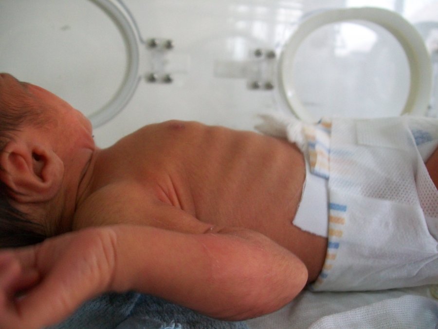
Not all people with neonatal Alagille syndrome have the same symptoms present. The condition can just cause minor issues like some changes to facial features, or it can cause severe heart and liver disease. Some of the most common symptoms include:

  • Deep-set eyes
  • Straight nose
  • Triangular face shape with pointed chin
  • Jaundice
  • Heart murmurs
  • Cardiac anomaly
  • Short height
  • Butterfly shaped vertebrae
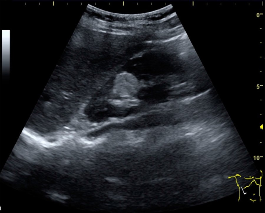
  • Kidney abnormalities
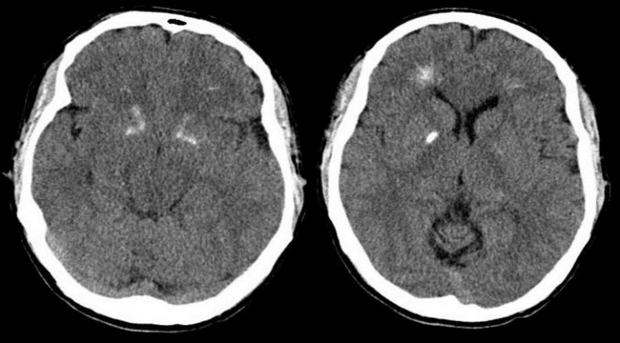
  • Eye abnormalities

Parents should keep in mind that some symptoms, like kidney abnormalities, can take a while to notice. Signs that these organs are not functioning properly can include chronic itchiness, fatigue, pale skin, fluid retention, weakness, or abnormally pale feces.

How Is Neonatal Alagille Syndrome Treated?

Treatment for neonatal alagille syndrome typically relies on what symptoms the patient is presenting. Many children have mild enough Alagille syndrome that they just need to eat a healthy diet and get plenty of rest to help with issues like delayed puberty or failure to thrive. Certain medications can help with things like improving bile function, stabilizing kidney function, and helping the heart run properly. However, in more serious situations, surgery may be necessary. Surgeries can help to correct skeletal issues, replace malfunctioning kidneys and livers, or treat cardiac abnormalities.

dr-allen-cherer-echogenic

Echogenic Bowel: An Overview

Though it is not an incredibly common diagnosis, there are still thousands of fetuses diagnosed with an echogenic bowel each year. Roughly 1.8 percent of all fetuses have echogenic bowel, and their parents often wonder what exactly this term means. Understanding all the details about echogenic bowel will help parents discover how to handle this condition.

What Is Echogenic Bowel?

This term is simply used to refer to a fetal bowel that is abnormally bright when viewed on an ultrasound. Ultrasounds are always in shades of black, grey, and white, with denser tissue being bright white and fluid being black. Usually, the bowels of the fetus tend to be a dark grey on an ultrasound because they are softened tissue, but if a fetus has echogenic bowel, their bowels may look as bright as thick bones like the pelvic bone.

What Does It Mean to Have Echogenic Bowel?

When parents first hear this word, they often start to wonder if it is a serious problem. The reality is that it is not always a sign of a health condition. 0.5 percent of all perfectly healthy fetuses have an echogenic bowel, and it can just occur due to various fluctuations in growth. However, echogenic bowel can be a cause for concern because it is more common in babies born with Down syndrome or cystic fibrosis. It can also be a sign that the fetus is suffering from an intestinal obstruction or an infection like cytomegalovirus or toxoplasmosis.

How Is Echogenic Bowel Treated?

The method for addressing echogenic bowel typically depends on the severity. Low grade echogenic bowels which are less dense than bone are normally harmless, so doctors tend to take a “wait and see” approach. They normally recommend taking a more detailed ultrasound in a few weeks to check up on the growth of the fetus. However, for more severely echogenic bowels, doctors may recommend a maternal serum screening, blood tests, or an amniocentesis to determine if the fetus has Down syndrome, infections, or cystic fibrosis. To make sure that the pregnancy continues safely, doctors will usually do regular fetal monitoring to make sure the fetus is growing properly following a display of echogenic bowel.

dr-allen-cherer-syndrome

A Closer Look at Prader-Willi Syndrome

Prader-Willi syndrome is a genetic disorder that is generally rare. It occurs in approximately one out of every 12,000 births and results in weak muscle tone, delayed development, and poor growth. Around the age of two, children with the condition develop an insatiable appetite that can lead to obesity. The condition was first identified by Drs. Labhart, Prader, and Willi in 1965.

Symptoms

Symptoms of Prader-Willi syndrome include:

  • Weak muscle tone
  • Poor feeding in newborns
  • Delayed development
  • Obesity
  • Chronic overeating (hyperphagia)
  • Distinct facial features, which include almond-shaped eyes, turned-down mouth, narrowing of the forehead at the temples and a thin upper lip
  • Underdeveloped genitals
  • Compulsive behavior
  • Extreme stubbornness
  • Angry outbursts
  • Short stature
  • Delayed puberty
  • Sleep problems

Cause

Prader-Willi syndrome is caused by a loss of the function of the genes 11-13 on chromosome 15. In an estimated 70 to 80 percent of people with the syndrome, the region of genes is missing due to deletion of paternal genes. In other cases, there may be two copies of chromosome 15 inherited from the mother or a defect in the paternal genes on chromosome 15.

The loss of function on chromosome 15 disrupts the operation of a part of the brain known as the hypothalamus. The hypothalamus is responsible for the release of hormones in the body. Without the normal release of hormones, growth, body temperature, hunger, sleep, and sexual development can all be affected.

Complications

Obesity is common in those with Prader-Willi syndrome. People with the condition have lower muscle mass and are less likely to be physically active. As a result, they need fewer calories. Unfortunately, the hypothalamus is responsible for releasing hormones to signal feelings of fullness after eating. This results in people with the condition eating too many calories and becoming overweight or extremely obese.

Type 2 diabetes, sleep apnea, gallbladder, heart and liver problems may all result from obesity. People with the condition are encouraged to work with a dietitian to eat the proper amount of calories in order to prevent complications from occurring. Additionally, most people with Prader-Willi syndrome may experience fertility problems and are at an increased likelihood of developing osteoporosis.

dr-allen-cherer-brain

DiGeorge Syndrome: An Overview

Primarily observed in children under the age of two, DiGeorge Syndrome is a rare neonatal chromosomal disorder affecting bodily development. The condition, which is also called 22q11.2 deletion syndrome, is caused by a defect in chromosome 22 and has a varying range of symptoms impacting both physical and mental growth. In some cases, it can be passed genetically from an affected parent to a child.

Due to the condition’s long list of symptoms — many of which are caused by a myriad of other conditions — DiGeorge Syndrome can be difficult to diagnose and treat, which means you will need to seek out an accurate and prompt evaluation from a trusted healthcare professional. Starting out, however, here is a quick overview of the condition to potentially point you in the right direction.

Knowing the signs

As mentioned before, symptoms of DiGeorge Syndrome can be vast, and therefore the condition can be hard to pinpoint at first. That said, there are several broad symptoms that have become listed as common warning signs; these include, but are certainly not limited to:

  • Cyanosis (a bluish tint to the skin caused by a lack of oxygen).
  • Learning difficulties, including those typically associated with Attention Deficit Disorder (ADD)
  • Skeletal abnormalities
  • Seizures and other epileptic symptoms
  • Feeding problems and failure to gain weight

Other, less externally evident symptoms may include autoimmune disorders, heart murmurs, frequent infections, and issues with the palate. In any scenario, the easiest and most established way to diagnose the condition is genetic testing, which can

Treating DiGeorge Syndrome

Unfortunately, there is currently no known cure for DiGeorge Syndrome — though certain symptoms may be individually treatable; this can fluctuate based on the urgency of the symptoms in question. For example, symptoms associated with certain immune-based disorders should ideally be addressed sooner than later to increase the chances of effective treatment. Other condition-based byproducts, like learning disabilities and anxiety, can be mitigated through proper intervention aimed at fostering intellectual growth and emotional stability.

These efforts may not treat the underlying root of the issue, but they can help on other fronts, such as improving the child’s overall quality of life.

Prevention can also be key in stopping the condition; if you feel you may have a family history of DiGeorge Syndrome, consult a specialist prior to any future pregnancies.

dr-allen-cherer-distress

Respiratory Distress in Newborns

Respiratory distress syndrome, or RDS, is a common lung complaint for infants. This is especially true in premature babies, born before 37 weeks. The more premature the baby, the greater the chance the child will develop RDS.

RDS is caused by a shortage of pulmonary surfactant. Surfactant is a liquid that helps keep air sacs in the lungs, known as alveoli, open. Alveoli are critical. They are the site of the exchange of oxygen and carbon dioxide. They make it possible for the blood to be oxygenated fully. Since surfactant makes this possible, it’s a very important substance indeed.

There are several risk factors for RDS. In addition to prematurity, babies with RDS are more likely to be white, male, and multiples. Mothers with diabetes are more likely to give birth to RDS babies. Babies delivered by c-section are also more likely to develop this problem.

Parents of babies with some of these risk factors should be aware of the symptoms of respiratory distress syndrome. Babies with RDS breathe fast. They may grunt, making an ugh sound with each breath. Their nostrils will flare every time they breathe. Finally, they can have retractions, where the skin pulls under the rib cage or in between ribs with each breath. Their skin may not be as pink as that of a typical baby.

Luckily, there is treatment for RDS these days. Delivery of oxygen by nasal cannula is one treatment. A CPAP, or continuous positive airway pressure, machine can be used to push air into an infant’s lungs. This will keep the alveoli open. Severe cases of RDS can require a ventilator.

Ventilators are a serious measure. They require intubation, or a tube being placed down the infant’s windpipe. Ventilators are only used in babies who can’t breathe well without assistance.

In addition to helping deliver more oxygen, the issue of a lack of surfactant can also be addressed. Surfactant can be delivered directly to the lungs, also via intubation. Medications to calm the infant are also used, especially when intubation is required.

RDS can sometimes also be associated with infections. In those cases, antibiotics may be given to the infant. Not every baby requires all of these treatments. In some cases, babies get worse before getting better. RDS is, in general, very treatable.

Powered by WordPress & Theme by Anders Norén